Comparison of Different Methods for Tissue Segmentation in Histopathological Whole-Slide Images
Publication
Metrics
AI Quick Summary
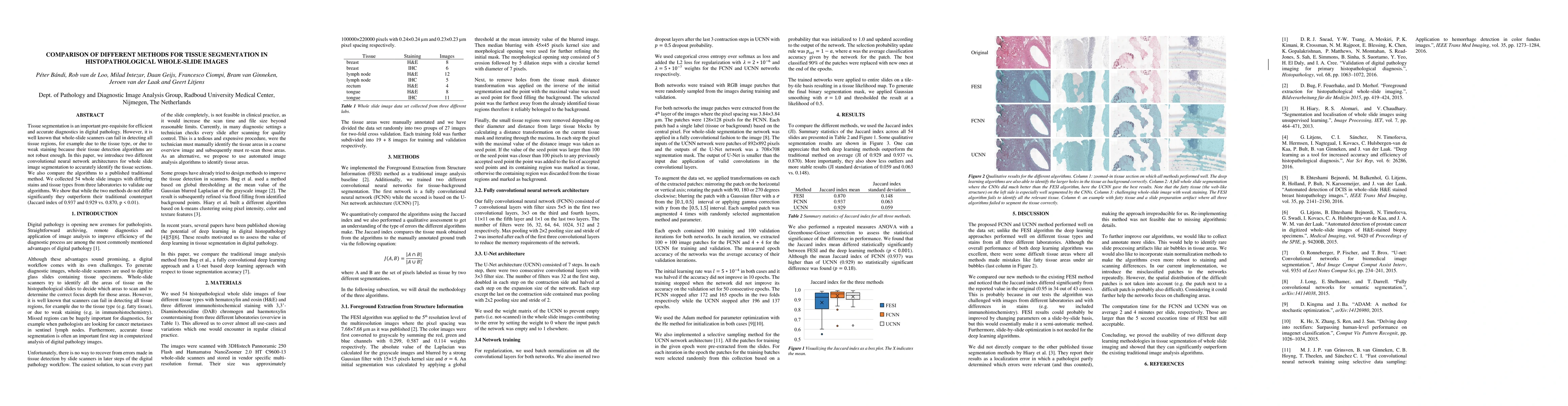
This paper compares two convolutional neural network architectures for tissue segmentation in histopathological whole-slide images, demonstrating their superior performance over a traditional method (Jaccard index of 0.937 and 0.929 vs. 0.870, p < 0.01). The study validates the algorithms using 54 diverse images from three laboratories.
Paper Preview
Abstract
Tissue segmentation is an important pre-requisite for efficient and accurate diagnostics in digital pathology. However, it is well known that whole-slide scanners can fail in detecting all tissue regions, for example due to the tissue type, or due to weak staining because their tissue detection algorithms are not robust enough. In this paper, we introduce two different convolutional neural network architectures for whole slide image segmentation to accurately identify the tissue sections. We also compare the algorithms to a published traditional method. We collected 54 whole slide images with differing stains and tissue types from three laboratories to validate our algorithms. We show that while the two methods do not differ significantly they outperform their traditional counterpart (Jaccard index of 0.937 and 0.929 vs. 0.870, p < 0.01).
AI Key Findings
Get AI-generated insights about this paper's methodology, results, significance, and more — seven facets brought into focus.
Impact
Paper Details
PDF Preview
Key Terms
Citation Network
Current paper (gray), citations (green), references (blue)
Display is limited for performance on very large graphs.
Discussion 0