Summary
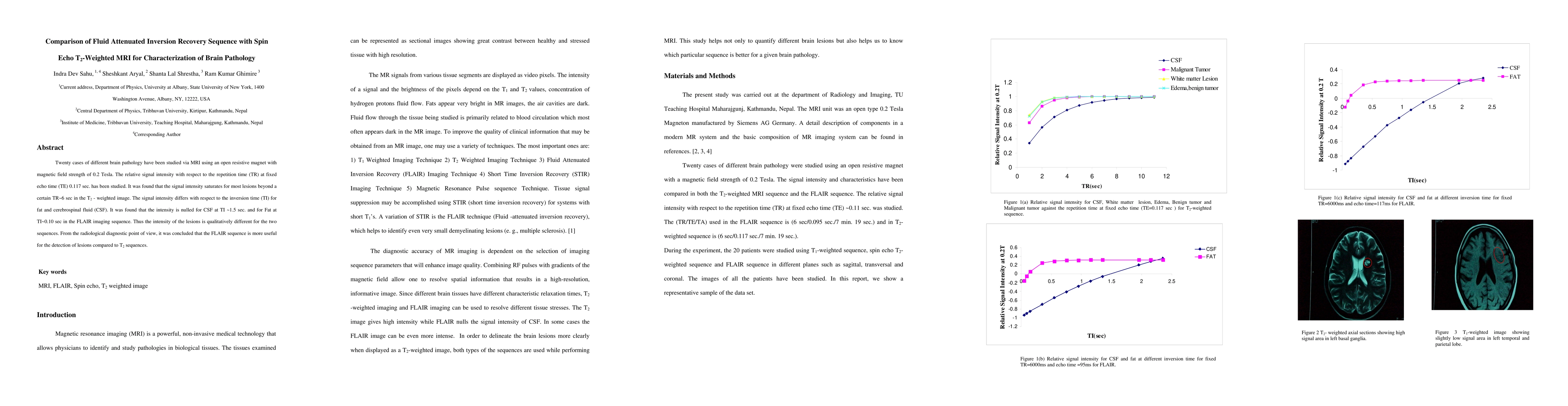
Twenty cases of different brain pathology have been studied via MRI using an open resistive magnet with magnetic field strength of 0.2 Tesla. The relative signal intensity with respect to the repetition time (TR) at fixed echo time (TE) 0.117 sec. has been studied. It was found that the signal intensity saturates for most lesions beyond a certain TR~6 sec in the T2 - weighted image. The signal intensity differs with respect to the inversion time (TI) for fat and cerebrospinal fluid (CSF). It was found that the intensity is nulled for CSF at TI ~1.5 sec. and for Fat at TI~0.10 sec in the FLAIR imaging sequence. Thus the intensity of the lesions is qualitatively different for the two sequences. From the radiological diagnostic point of view, it was concluded that the FLAIR sequence is more useful for the detection of lesions compared to T2 sequences.
AI Key Findings
Get AI-generated insights about this paper's methodology, results, and significance.
Paper Details
PDF Preview
Key Terms
Citation Network
Current paper (gray), citations (green), references (blue)
Display is limited for performance on very large graphs.
Similar Papers
Found 4 papersTemporally Adjustable Longitudinal Fluid-Attenuated Inversion Recovery MRI Estimation / Synthesis for Multiple Sclerosis
Jueqi Wang, Jacob Levman, Derek Berger et al.
T2 mapping at 0.55 T using Ultra-Fast Spin Echo MRI
Meritxell Bach Cuadra, Tom Hilbert, Tobias Kober et al.
Triangular Fibrocartilage Characterization with Ultrashort Echo Time-T2* MRI: Insights from a Healthy Cohort.
Boudabbous, Sana, Poletti, Pierre-Alexandre, Bouredoucen, Hicham et al.
| Title | Authors | Year | Actions |
|---|
Comments (0)