This paper evaluates various U-net-based convolutional neural networks for liver segmentation in CT scans, finding that 2D slice-wise approaches achieve mean and median Dice coefficients above 0.97, outperforming 3D approaches despite current hardware constraints.
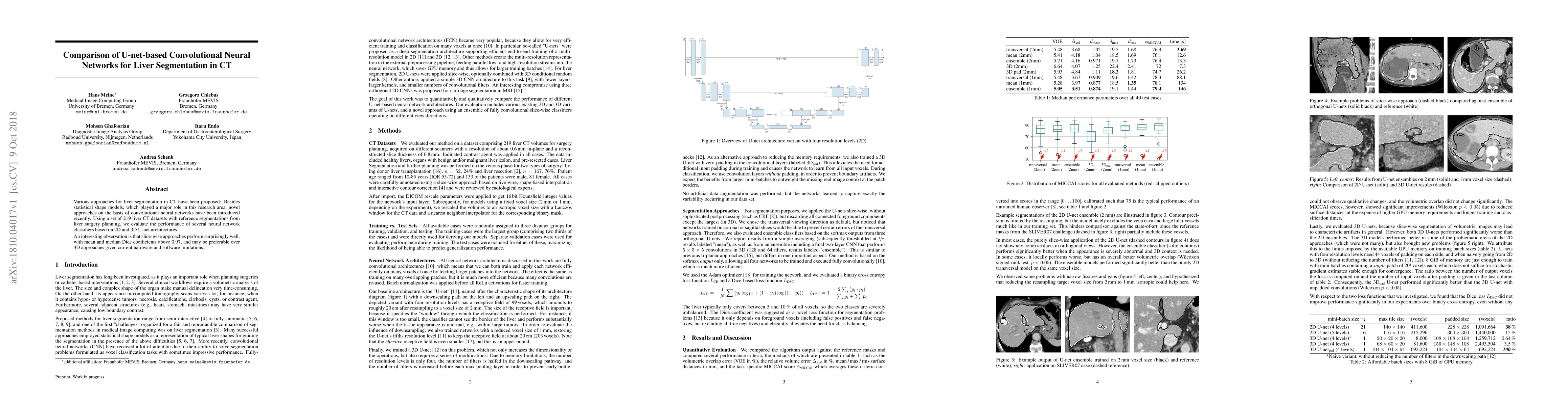
Various approaches for liver segmentation in CT have been proposed: Besides statistical shape models, which played a major role in this research area, novel approaches on the basis of convolutional neural networks have been introduced recently. Using a set of 219 liver CT datasets with reference segmentations from liver surgery planning, we evaluate the performance of several neural network classifiers based on 2D and 3D U-net architectures. An interesting observation is that slice-wise approaches perform surprisingly well, with mean and median Dice coefficients above 0.97, and may be preferable over 3D approaches given current hardware and software limitations.
Get AI-generated insights about this paper's methodology, results, and significance.
Current paper (gray), citations (green), references (blue)
Title | Authors | Year | Actions |
---|---|---|---|
Artificial Intelligence in Imaging of Chronic Liver Diseases: Current Update and Future Perspectives.
|
C. Sabottke, B. Spieler, A. Moawad, K. Elsayes
|
2021
|
View Paper |
Comparison of 2D vs 3D U-Net Organ Segmentation in abdominal 3D CT images
|
N. Zettler, André Mastmeyer
|
2021
|
View Paper |
Artificial intelligence in assessment of hepatocellular carcinoma treatment response
|
B. Spieler, C. Sabottke, A. Moawad, A. Gabr, M. Bashir, R. Do, V. Yaghmai, R. Rozenberg, M. Gerena, J. Yacoub, K. Elsayes
|
2021
|
View Paper |
2D vs. 3D U-Net abdominal organ segmentation in CT data using organ bounds
|
D. Kern, U. Klauck, T. Ropinski, André Mastmeyer
|
2021
|
View Paper |
Automatic Skull Stripping of Rat and Mouse Brain MRI Data Using U-Net
|
Li-Ming Hsu, Shuai Wang, Paridhi Ranadive, Woomi Ban, T. Chao, Shengdeng Song, Domenic H. Cerri, Lindsay R. Walton, Margaret A. Broadwater, Sung-Ho Lee, D. Shen, Y. Shih
|
2020
|
View Paper |
Liver Segmentation on a Variety of Computed Tomography (CT) Images Based on Convolutional Neural Networks Combined with Connected Components
|
Hoang Hong Son, P. C. Phuong, T. Walsum, Luu Manh Ha
|
2020
|
View Paper |
Automatic Multimodal Registration via Intraprocedural Cone-Beam CT Segmentation using MRI Distance Maps
|
Z. Augenfeld, Ming De Lin, J. Chapiro, J. Duncan
|
2020
|
View Paper |
Automatic multi-object organ detection and segmentation in abdominal CT data
|
Oliver Mietzner, André Mastmeyer
|
2020
|
View Paper |
An Evaluation of CNN-based Liver Segmentation Methods using Multi-types of CT Abdominal Images from Multiple Medical Centers
|
Hoang Hong Son, Cam Phuong Pham, D. Franklin, T. Walsum, H. M. Luu
|
2019
|
View Paper |
A Study on the Liver and Tumor Segmentation and Hologram Visualization of CT Images Using Deep Learning
|
†. DaeJinKim, Y. J. Kim, Y. Jeon, T. Hwang, Seok-Won Choi, J. Baek, K. Kim
|
2022
|
View Paper |
Pre- and Post-processing Strategies for Generic Slice-wise Segmentation of Tomographic 3D Datasets Utilizing U-Net Deep Learning Models Trained for Specific Diagnostic Domains
|
G. Zwettler, W. Backfrieder, D. Holmes
|
2020
|
View Paper |
An uncertainty-aware deep learning architecture with outlier An uncertainty-aware deep learning architecture with outlier mitigation for prostate gland segmentation in radiotherapy mitigation for prostate gland segmentation in radiotherapy treatment planning treatment planning
|
Xin Li, H. Bagher-Ebadian, Stephen Gardner, Joshua Kim, Mohamed Elshaikh, B. Movsas, Dongxiao Zhu, I. Chetty
|
View Paper |
Comments (0)