Publication
Metrics
AI Quick Summary
This paper proposes a novel complementary speckle STED microscopy technique using speckle patterns to enhance spatial resolution and reduce photobleaching, demonstrating improved temporal resolution and effective three-dimensional imaging in biological samples.
Paper Preview
Abstract
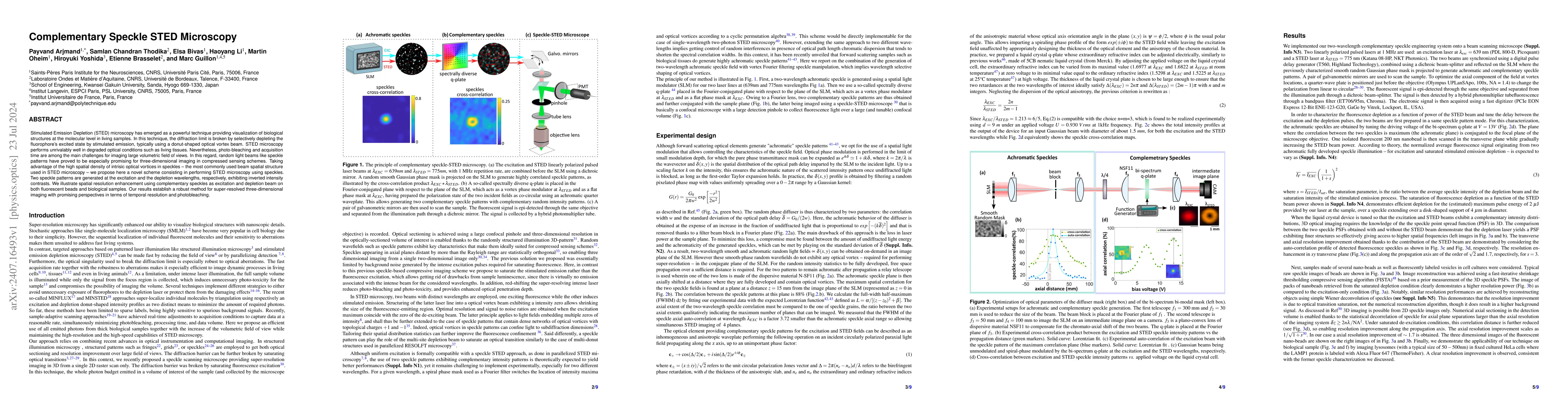
Stimulated Emission Depletion (STED) microscopy has emerged as a powerful technique providing visualization of biological structures at the molecular level in living samples. In this technique, the diffraction limit is broken by selectively depleting the fluorophore's excited state by stimulated emission, typically using a donut-shaped optical vortex beam. STED microscopy performs unrivalably well in degraded optical conditions such as living tissues. Nevertheless, photo-bleaching and acquisition time are among the main challenges for imaging large volumetric field of views. In this regard, random light beams like speckle patterns have proved to be especially promising for three-dimensional imaging in compressed sensing schemes. Taking advantage of the high spatial density of intrisic optical vortices in speckles -- the most commonly used beam spatial structure used in STED microscopy -- we propose here a novel scheme consisting in performing STED microscopy using speckles. Two speckle patterns are generated at the excitation and the depletion wavelengths, respectively, exhibiting inverted intensity contrasts. We illustrate spatial resolution enhancement using complementary speckles as excitation and depletion beam on both fluorescent beads and biological samples. Our results establish a robust method for super-resolved three-dimensional imaging with promising perspectives in terms of temporal resolution and photobleaching.
AI Key Findings
Get AI-generated insights about this paper's methodology, results, significance, and more — seven facets brought into focus.
Impact
Authors
PDF Preview
Citation Network
Current paper (gray), citations (green), references (blue)
Display is limited for performance on very large graphs.
Discussion 0