Complementary structural and functional abnormalities to localise epileptogenic tissue
Publication
Metrics
AI Quick Summary
This study investigates the use of structural connectivity abnormalities from diffusion-weighted MRI (dMRI) alongside functional abnormalities from intracranial EEG (iEEG) to better localize the epileptogenic zone and improve surgical outcomes in drug-refractory focal epilepsy. Combining both modalities significantly enhanced the prediction of seizure freedom post-surgery.
Paper Preview
Abstract
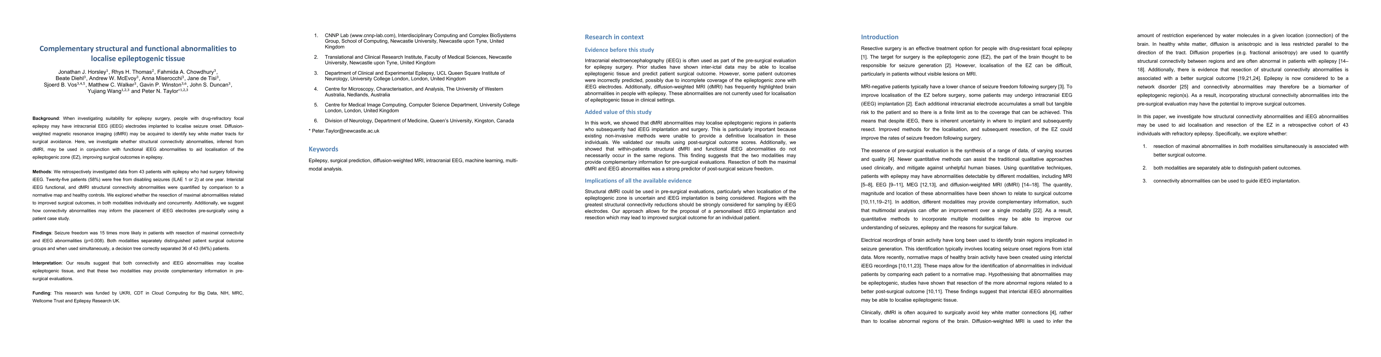
When investigating suitability for surgery, people with drug-refractory focal epilepsy may have intracranial EEG (iEEG) electrodes implanted to localise seizure onset. Diffusion-weighted magnetic resonance imaging (dMRI) may be acquired to identify key white matter tracts for surgical avoidance. Here, we investigate whether structural connectivity abnormalities, inferred from dMRI, may be used in conjunction with functional iEEG abnormalities to aid localisation and resection of the epileptogenic zone (EZ), and improve surgical outcomes in epilepsy. We retrospectively investigated data from 43 patients with epilepsy who had surgery following iEEG. Twenty five patients (58%) were free from disabling seizures (ILAE 1 or 2) at one year. For all patients, T1-weighted and diffusion-weighted MRIs were acquired prior to iEEG implantation. Interictal iEEG functional, and dMRI structural connectivity abnormalities were quantified by comparison to a normative map and healthy controls respectively. First, we explored whether the resection of maximal (dMRI and iEEG) abnormalities related to improved surgical outcomes. Second, we investigated whether the modalities provided complementary information for improved prediction of surgical outcome. Third, we suggest how dMRI abnormalities may be useful to inform the placement of iEEG electrodes as part of the pre-surgical evaluation using a patient case study. Seizure freedom was 15 times more likely in those patients with resection of maximal dMRI and iEEG abnormalities (p=0.008). Both modalities were separately able to distinguish patient outcome groups and when combined, a decision tree correctly separated 36 out of 43 (84%) patients based on surgical outcome. Structural dMRI could be used in pre-surgical evaluations, particularly when localisation of the EZ is uncertain, to inform personalised iEEG implantation and resection.
AI Key Findings
Get AI-generated insights about this paper's methodology, results, significance, and more — seven facets brought into focus.
Impact
Paper Details
Authors
PDF Preview
Key Terms
Citation Network
Current paper (gray), citations (green), references (blue)
Display is limited for performance on very large graphs.
Discussion 0