ArxivLens

Quick Summary:

This research classifies over 180,000 protein-ligand binding sites into approximately 3000 structural motifs through atomic-level comparisons, revealing that most motifs are unique to specific protein families and associated with particular ligands, while also identifying over 4000 shared binding site pairs across different protein folds.

Journal Reference: Structure, 17:234-246 (2009)
DOI: 10.1016/j.str.2008.11.009
arXiv Id: 0810.1327
Comments: 13 pages, 8 figures
Date Published: 2009-02-11
Date Updated: 2009-02-11

Categories:

Key Terms (TF-IDF):

protein (0.394)
sites (0.312)
binding (0.306)
structural (0.247)
comparison (0.171)
structures (0.160)
recently developed (0.150)
universality (0.139)
designing (0.138)
confined (0.135)
database (0.133)
shared (0.132)
alignment (0.130)
diversity (0.129)
families (0.127)
engineering (0.126)
similarity (0.126)
approximately (0.122)
activity (0.121)
studying (0.120)
License: http://arxiv.org/licenses/nonexclusive-distrib/1.0/

Summary:

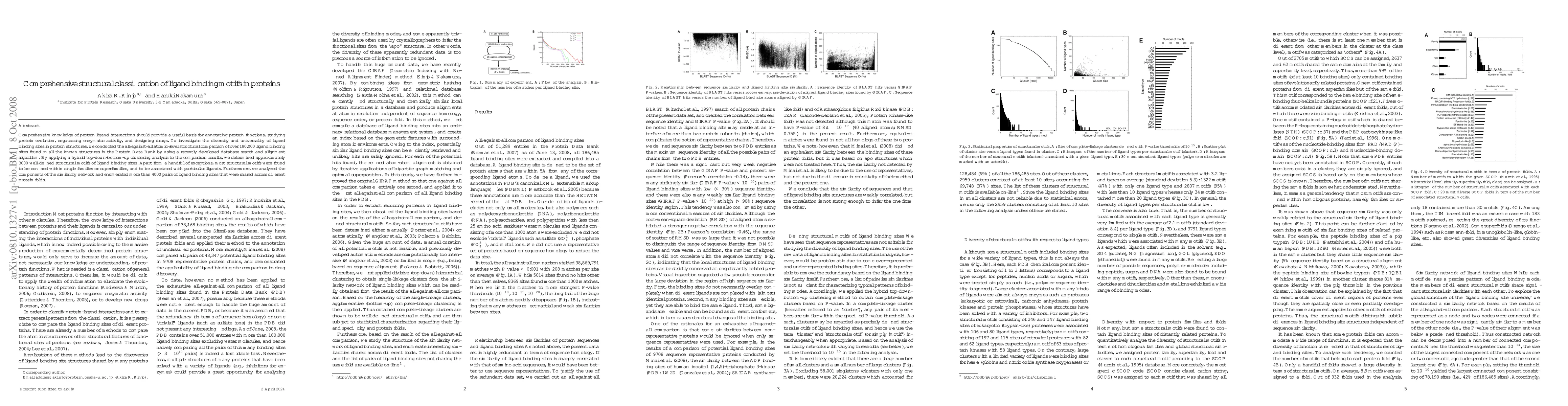
Comprehensive knowledge of protein-ligand interactions should provide a useful basis for annotating protein functions, studying protein evolution, engineering enzymatic activity, and designing drugs. To investigate the diversity and universality of ligand binding sites in protein structures, we conducted the all-against-all atomic-level structural comparison of over 180,000 ligand binding sites found in all the known structures in the Protein Data Bank by using a recently developed database search and alignment algorithm. By applying a hybrid top-down-bottom-up clustering analysis to the comparison results, we determined approximately 3000 well-defined structural motifs of ligand binding sites. Apart from a handful of exceptions, most structural motifs were found to be confined within single families or superfamilies, and to be associated with particular ligands. Furthermore, we analyzed the components of the similarity network and enumerated more than 4000 pairs of ligand binding sites that were shared across different protein folds.

Citation & Reference Network Visualization

This graph shows how this paper is connected to other research. The current paper is shown in dark gray, papers that cite it are in green, and papers it references are in blue. You can:

Current Paper
Papers citing this work (Citations)
Papers cited by this work (References)

• Hover over any node to see the paper title

• Click a node to view that paper's details

• Drag nodes to rearrange the graph

• Use the zoom controls (bottom right) to zoom in/out

Citations and References

Title Authors Year Actions
DEPACT and PACMatch: A Workflow of Designing De Novo Protein Pockets to Bind Small Molecules
Yaoxi Chen, Quan Chen, Haiyan Liu
2022
View Paper
Defining A Global Map of Functional Group-based 3D Ligand-binding Motifs
Liu Yang, Wei He, Y. Yun, Yongxiang Gao, Zhong-liang Zhu, M. Teng, Zhi Liang, L. Niu
2020
View Paper
BERTology Meets Biology: Interpreting Attention in Protein Language Models
Jesse Vig, Ali Madani, L. Varshney, Caiming Xiong, R. Socher, Nazneen Rajani
2020
View Paper
DeepDrug3D: Classification of ligand-binding pockets in proteins with a convolutional neural network
Limeng Pu, R. Govindaraj, J. Lemoine, Hsiao-Chun Wu, Michal Brylinski
2019
View Paper
Universal Architectural Concepts Underlying Protein Folding Patterns
A. Lesk, R. Subramanian, L. Allison, David Abramson, Peter James Stuckey, M. G. D. L. Banda, A. S. Konagurthu
2018
View Paper
Ligand Binding Site Structure Influences the Evolution of Protein Complex Function and Topology
György Abrusán, J. Marsh
2018
View Paper
An Augmented Pocketome: Detection and Analysis of Small-Molecule Binding Pockets in Proteins of Known 3D Structure.
Raghu Bhagavat, Santhosh Sankar, N. Srinivasan, N. Chandra
2018
View Paper
Global organization of a binding site network gives insight into evolution and structure-function relationships of proteins
Juyong Lee, Janez Konc, D. Janežič, B. Brooks
2017
View Paper
Cooperative "folding transition" in the sequence space facilitates function-driven evolution of protein families.
A. Kinjo
2017
View Paper
The pimeloyl-CoA synthetase BioW defines a new fold for adenylate-forming enzymes.
P. Estrada, Miglena Manandhar, S. Dong, J. Deveryshetty, V. Agarwal, J. Cronan, S. Nair
2017
View Paper
LibME—automatic extraction of 3D ligand‐binding motifs for mechanistic analysis of protein–ligand recognition
Wei He, Zhi Liang, M. Teng, L. Niu
2016
View Paper
Protein Data Bank Japan (PDBj): updated user interfaces, resource description framework, analysis tools for large structures
A. Kinjo, G. Bekker, Hirofumi Suzuki, Y. Tsuchiya, T. Kawabata, Yasuyo Ikegawa, Haruki Nakamura
2016
View Paper
Landscape of protein–small ligand binding modes
K. Kasahara, K. Kinoshita
2016
View Paper
Impact of Binding Site Comparisons on Medicinal Chemistry and Rational Molecular Design.
C. Ehrt, Tobias Brinkjost, O. Koch
2016
View Paper
Considerations of Protein Subpockets in Fragment‐Based Drug Design
Matthew D. Bartolowits, V. Davisson
2016
View Paper
Many InChIs and quite some feat
W. Warr
2015
View Paper
Protein function from its emergence to diversity in contemporary proteins
A. Goncearenco, I. Berezovsky
2015
View Paper
Systematic investigation of sequence and structural motifs that recognize ATP
Ke Chen, Dacheng Wang, Lukasz Kurgan
2015
View Paper
PoSSuM v.2.0: data update and a new function for investigating ligand analogs and target proteins of small-molecule drugs
J. Ito, K. Ikeda, Kazunori D. Yamada, K. Mizuguchi, K. Tomii
2014
View Paper
AFAL: a web service for profiling amino acids surrounding ligands in proteins
Mauricio Arenas-Salinas, S. Ortega-Salazar, Fernando Gonzales-Nilo, E. Pohl, D. Holmes, Raquel Quatrini
2014
View Paper
PLIC: protein–ligand interaction clusters
Praveen Anand, D. Nagarajan, S. Mukherjee, N. Chandra
2014
View Paper
Protein Binding Site Similarities as Driver for Drug Repositioning
J. Haupt
2014
View Paper
Identification of Similar Binding Sites to Detect Distant Polypharmacology
Xavier Jalencas, J. Mestres
2013
View Paper
Exhaustive comparison and classification of ligand-binding surfaces in proteins
Yoichi Murakami, K. Kinoshita, A. Kinjo, Haruki Nakamura
2013
View Paper
IMAAAGINE: a webserver for searching hypothetical 3D amino acid side chain arrangements in the Protein Data Bank
Nurul Nadzirin, P. Willett, P. Artymiuk, M. F. Raih
2013
View Paper
MICAN : a protein structure alignment algorithm that can handle Multiple-chains, Inverse alignments, Cα only models, Alternative alignments, and Non-sequential alignments
S. Minami, Kengo Sawada, G. Chikenji
2013
View Paper
Conservation of water molecules in protein binding interfaces
Zhenhua Li, Ying He, Longbing Cao, L. Wong, Jinyan Li
2012
View Paper
GIRAF: a method for fast search and flexible alignment of ligand binding interfaces in proteins at atomic resolution
A. Kinjo, Haruki Nakamura
2012
View Paper
SPRITE and ASSAM: web servers for side chain 3D-motif searching in protein structures
Nurul Nadzirin, Eleanor J. Gardiner, P. Willett, P. Artymiuk, M. F. Raih
2012
View Paper
PDB‐scale analysis of known and putative ligand‐binding sites with structural sketches
J. Ito, Yasuo Tabei, Kana Shimizu, K. Tomii, K. Tsuda
2012
View Paper
Protein Data Bank Japan (PDBj): maintaining a structural data archive and resource description framework format
A. Kinjo, Hirofumi Suzuki, R. Yamashita, Yasuyo Ikegawa, Takahiro Kudou, Reiko Igarashi, Yumiko Kengaku, Hasumi Cho, D. Standley, A. Nakagawa, Haruki Nakamura
2011
View Paper
Protein Data Bank Japan (PDBj): an interview with Haruki Nakamura of Osaka University
W. Warr
2011
View Paper
Composite Structural Motifs of Binding Sites for Delineating Biological Functions of Proteins
A. Kinjo, Haruki Nakamura
2011
View Paper
Prediction of ligand‐binding sites of proteins by molecular docking calculation for a random ligand library
Y. Fukunishi, Haruki Nakamura
2011
View Paper
Knowledge-based annotation of small molecule binding sites in proteins
Ratna R. Thangudu, M. Tyagi, Benjamin A. Shoemaker, S. Bryant, A. Panchenko, T. Madej
2010
View Paper
Geometric similarities of protein-protein interfaces at atomic resolution are only observed within homologous families: an exhaustive structural classification study.
A. Kinjo, Haruki Nakamura
2010
View Paper
Analysis of substructural variation in families of enzymatic proteins with applications to protein function prediction
D. Bryant, Mark Moll, B. Chen, V. Fofanov, L. Kavraki
2010
View Paper
Protein Pockets: Inventory, Shape, and Comparison
R. Coleman, K. Sharp
2010
View Paper
A hybrid clustering of protein binding sites
Gábor Iván, Zoltan Szabadka, V. Grolmusz
2010
View Paper
Artificial Intelligence in Medicine - 18th International Conference on Artificial Intelligence in Medicine, AIME 2020, Minneapolis, MN, USA, August 25-28, 2020, Proceedings
Martin Michalowski, Robert Moskovitch, R. Goebel, Yuzuru Tanaka
2020
View Paper
Unsupervised Grammar Induction for Revealing the Internal Structure of Protein Sequence Motifs
O. Unold, M. Gábor, W. Dyrka
2020
View Paper
Identifying Functional Relationships Via the Annotation and Comparison of Three-Dimensional Amino Acid Arrangements in Protein Structures
M. F. Raih, N. S. A. Ghani
2019
View Paper
Discovery of molecules that modulate protein-protein interactions in the context of human proliferating cell nuclear antigen-associated processes of DNA replication and damage repair
Matthew D. Bartolowits
2015
View Paper
Functional structural motifs for protein-ligand, protein-protein, and protein-nucleic acid interactions and their connection to supersecondary structures.
A. Kinjo, Haruki Nakamura
2013
View Paper
Redundancy-aware learning of protein structure-function relationships
D. Bryant
2012
View Paper
Mapping the Structural Landscape of Protein Families with Geometric Feature Vectors by
L. Kavraki
2009
View Paper
Shortest Geometric Paths Analysis in Structural Biology
R. Coleman
2009
View Paper
Conservation of Water Molecules in Protein Binding Interfaces Conservation of Water Molecules
Zhenhua Li, Ying He, Longbing Cao, Limsoon Wong, Jinyan Li
View Paper

Comments (0)

This website uses cookies to ensure you get the best experience. Learn more