Publication
Metrics
AI Quick Summary
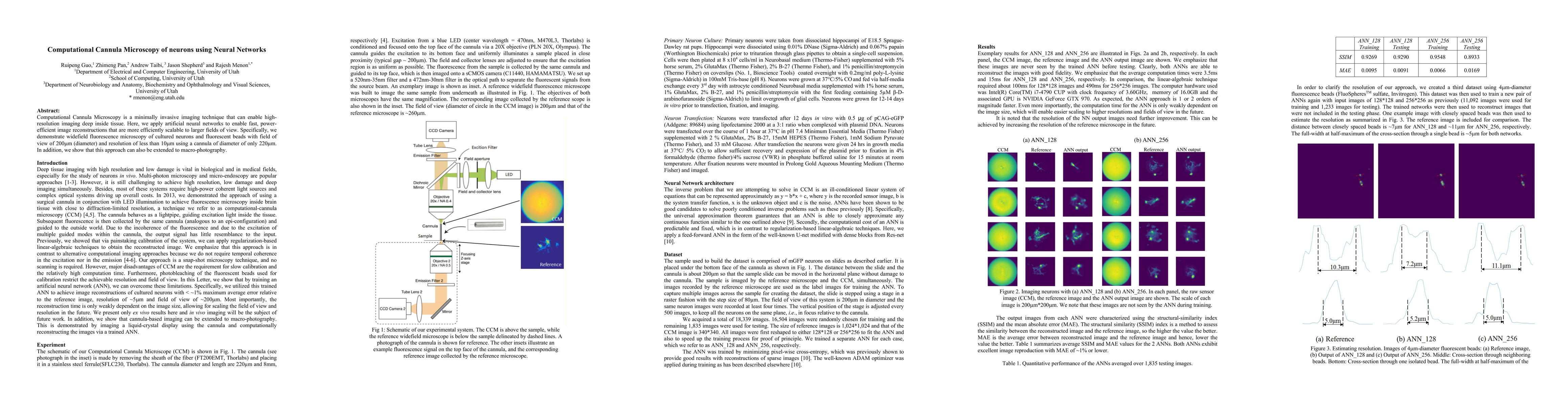
This paper introduces Computational Cannula Microscopy, a minimally invasive imaging technique that uses neural networks for fast, power-efficient image reconstructions. It demonstrates high-resolution imaging of neurons and fluorescent beads with a 200$\mu$m field of view and less than 10$\mu$m resolution through a 220$\mu$m cannula, and extends the method to macro-photography.
Paper Preview
Abstract
Computational Cannula Microscopy is a minimally invasive imaging technique that can enable high-resolution imaging deep inside tissue. Here, we apply artificial neural networks to enable fast, power-efficient image reconstructions that are more efficiently scalable to larger fields of view. Specifically, we demonstrate widefield fluorescence microscopy of cultured neurons and fluorescent beads with field of view of 200$\mu$m (diameter) and resolution of less than 10$\mu$m using a cannula of diameter of only 220$\mu$m. In addition, we show that this approach can also be extended to macro-photography.
AI Key Findings
Get AI-generated insights about this paper's methodology, results, significance, and more — seven facets brought into focus.
Impact
Paper Details
Authors
PDF Preview
Key Terms
Citation Network
Current paper (gray), citations (green), references (blue)
Display is limited for performance on very large graphs.
Discussion 0