This paper reviews current computational pathology methods for analyzing tissue data, highlighting challenges in integrating diverse data sources, and discusses potential future research directions to enhance diagnostic accuracy and treatment strategies in cancer detection.
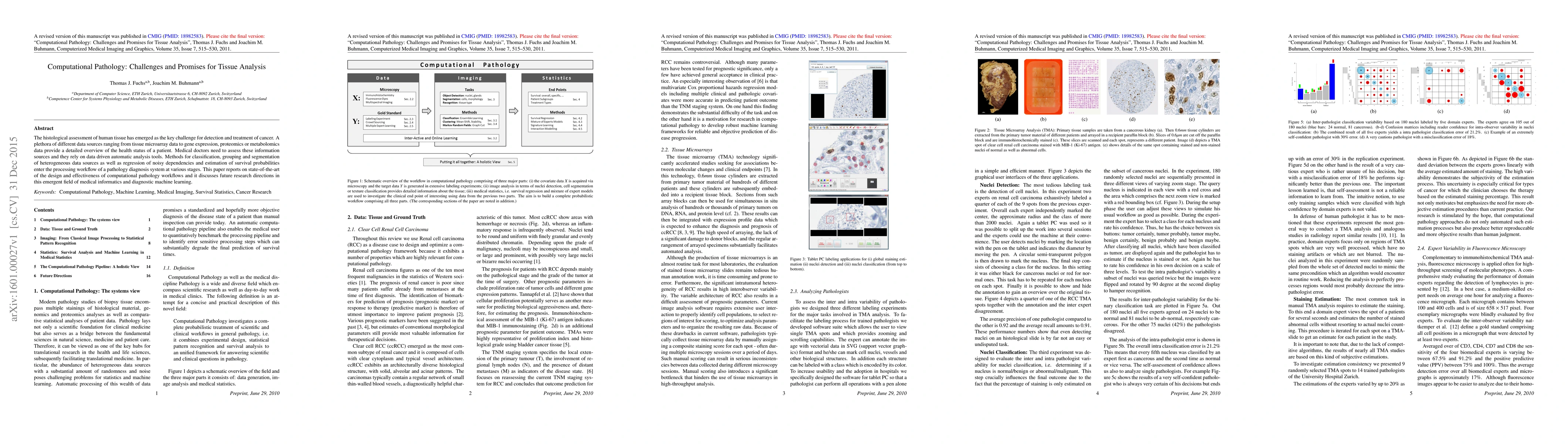
The histological assessment of human tissue has emerged as the key challenge for detection and treatment of cancer. A plethora of different data sources ranging from tissue microarray data to gene expression, proteomics or metabolomics data provide a detailed overview of the health status of a patient. Medical doctors need to assess these information sources and they rely on data driven automatic analysis tools. Methods for classification, grouping and segmentation of heterogeneous data sources as well as regression of noisy dependencies and estimation of survival probabilities enter the processing workflow of a pathology diagnosis system at various stages. This paper reports on state-of-the-art of the design and effectiveness of computational pathology workflows and it discusses future research directions in this emergent field of medical informatics and diagnostic machine learning.
Get AI-generated insights about this paper's methodology, results, and significance.
Current paper (gray), citations (green), references (blue)
Joel H. Saltz, Jakub R. Kaczmarzyk, Peter K. Koo
Nasir Rajpoot, Mohsin Bilal, Manahil Raza et al.
Liangrui Pan, Shaoliang Peng, Zhichao Feng
Title | Authors | Year | Actions |
---|---|---|---|
Enhancing HER2 testing in breast cancer: predicting fluorescence in situ hybridization (FISH) scores from immunohistochemistry images via deep learning
|
Daniel O Macaulay, Wenchao Han, Mark D. Zarella, Chris A Garcia, Thomas E. Tavolara
|
2025
|
View Paper |
VLEER: Vision and Language Embeddings for Explainable Whole Slide Image Representation
|
A. Nguyen, Keunho Byeon, Kyungeun Kim, Jin Tae Kwak
|
2025
|
View Paper |
A large expert-annotated single-cell peripheral blood dataset for hematological disease diagnostics
|
Sayedali Shetab Boushehri, A. Gruber, Salome Kazeminia, Christian Matek, Karsten Spiekermann, C. Pohlkamp, T. Haferlach, Carsten Marr
|
2025
|
View Paper |
CFI-ViT: A coarse-to-fine inference based vision transformer for gastric cancer subtype detection using pathological images
|
Xinghang Wang, Haibo Tao, Bin Wang, Huaiping Jin, Zhenhui Li
|
2025
|
View Paper |
Sparse vertex discriminant analysis: Variable selection for biomedical classification applications
|
Alfonso Landeros, Seyoon Ko, Jack Z. Chang, Tong Tong Wu, Kenneth Lange
|
2025
|
View Paper |
A GAN-Based Data Augmentation Method for Mitigating Class Imbalance Problem in Histopathological Image Classification
|
Lingling Yuan, M. Rahaman, Hongzan Sun, Chen Li, Yutong Gu, Tao Jiang, Marcin Grzegorzek, Xiaoyan Li
|
2024
|
View Paper |
A generalised vision transformer-based self-supervised model for diagnosing and grading prostate cancer using histological images
|
A. Chaurasia, H. C. Harris, P. W. Toohey, A. W. Hewitt
|
2024
|
View Paper |
Image Analysis in Histopathology and Cytopathology: From Early Days to Current Perspectives
|
Tibor Mezei, M. Kolcsár, András Joó, Simona Gurzu
|
2024
|
View Paper |
CNMI-YOLO: Domain Adaptive and Robust Mitosis Identification in Digital Pathology.
|
Yasemin Topuz, S. Yıldız, Songül Varlı
|
2024
|
View Paper |
Establishing Causal Relationship Between Whole Slide Image Predictions and Diagnostic Evidence Subregions in Deep Learning
|
Tianhang Nan, Yong Ding, Hao Quan, Deliang Li, Lisha Li, Guanghong Zhao, Xiaoyu Cui
|
2024
|
View Paper |
A foundation model for clinical-grade computational pathology and rare cancers detection
|
Eugene Vorontsov, A. Bozkurt, Adam Casson, George Shaikovski, Michal Zelechowski, Kristen Severson, Eric Zimmermann, James Hall, Neil Tenenholtz, Nicolò Fusi, Ellen Yang, Philippe Mathieu, A. van Eck, Donghun Lee, Julian Viret, Eric Robert, Yi Kan Wang, J. Kunz, Matthew C H Lee, Jan H Bernhard, R. Godrich, Gerard Oakley, Ewan Millar, Matthew G Hanna, Hannah Y Wen, Juan Retamero, William A. Moye, Razik Yousfi, C. Kanan, D.S. Klimstra, B. Rothrock, Siqi Liu, Thomas J Fuchs
|
2024
|
View Paper |
Histological Hyperspectral Glioblastoma Dataset (HistologyHSI-GB)
|
S. Ortega, Laura Quintana-Quintana, Raquel León, H. Fabelo, M. L. Plaza, R. Camacho, G. Callicó
|
2024
|
View Paper |
Towards non-invasive biomarkers in colorectal cancer management: A study on integrating radiomics and deep learning-based image processing for tumor-stroma interaction
|
P. E. Adjei, O. Otoo, Jinlang Yang, Nini Rao
|
2024
|
View Paper |
Computational pathology: an evolving concept
|
Ioannis Prassas, Blaise Clarke, Timothy Youssef, Juliana Phlamon, Lampros Dimitrakopoulos, Andrew Rofaeil, George M Yousef
|
2024
|
View Paper |
Cell-TIMP: Cellular Trajectory Inference based on Morphological Parameter
|
P. Raj, Himanshu Gupta, Pooja Anantha, Ishan Barman
|
2024
|
View Paper |
Machine learning-based pathomics signature of histology slides as a novel prognostic indicator in primary central nervous system lymphoma
|
Ling Duan, Yongqi He, Wenhui Guo, Yanru Du, Shuo Yin, Shoubo Yang, Gehong Dong, Wenbin Li, Feng Chen
|
2024
|
View Paper |
A histopathological image analysis for the classification of endocervical adenocarcinoma Silva patterns depend on weakly supervised deep learning.
|
Qingqing Liu, Xiaofang Zhang, Xuji Jiang, Chunyan Zhang, Jiamei Li, Xuedong Zhang, Jingyan Yang, Ning Yu, Yongcun Zhu, Jing Liu, Fengxiang Xie, Yawen Li, Yiping Hao, Yuan Feng, Qi Wang, Qun Gao, Wenjing Zhang, Teng Zhang, Taotao Dong, Baoxia Cui
|
2024
|
View Paper |
How cy pres promotes transdisciplinary convergence science: an academic health center for women’s cardiovascular and brain health
|
A. Villablanca, Brittany N. Dugger, Saivageethi Nuthikattu, Joohi Chauhan, Samson Cheung, Chen-Nee Chuah, Siedah L Garrison, D. Milenkovic, J. Norman, L. C. Oliveira, Bridgette P. Smith, Susan D. Brown
|
2024
|
View Paper |
Banff Digital Pathology Working Group: Image Bank, Artificial Intelligence Algorithm, and Challenge Trial Developments
|
A. Farris, Mariam P. Alexander, Ulysses Balis, Laura Barisoni, Peter Boor, R. Bülow, Lynn D. Cornell, A. Demetris, Evan Farkash, M. Hermsen, Julien Hogan, Renate Kain, J. Kers, Jun Kong, Richard M. Levenson, A. Loupy, M. Naesens, P. Sarder, John Tomaszewski, Jeroen van der Laak, Dominique van Midden, Yukako Yagi, Kim Solez
|
2023
|
View Paper |
Segmentation of Nuclei in H&E-Stained Histological Images using Deep Learning Framework: A Perspective on Ensemble Approach and Nuclei Count
|
Madhavi Aghera, Krishna Veer Singh, Kishankumar Vaishnani, Urvi Oza, Bakul Gohel
|
2023
|
View Paper |
Morphological diagnosis of hematologic malignancy using feature fusion-based deep convolutional neural network
|
D. P. Yadav, Deepak Kumar, Anand Singh Jalal, Ankit Kumar, K. Singh, Mohd Asif Shah
|
2023
|
View Paper |
Automatic Identification of Glomerular in Whole-Slide Images Using a Modified UNet Model
|
Gurjinder Kaur, Meenu Garg, Sheifali Gupta, Sapna Juneja, Junaid Rashid, Deepali Gupta, Asadullah Shah, A. Shaikh
|
2023
|
View Paper |
iQC: machine-learning-driven prediction of surgical procedure uncovers systematic confounds of cancer whole slide images in specific medical centers
|
A. J. Schaumberg, M. S. Lewis, R. Nazarian, A. Wadhwa, N. Kane, G. Turner, P. Karnam, P. Devineni, N. Wolfe, R. Kintner, M. Rettig, B. S. Knudsen, I. Garraway, S. Pyarajan
|
2023
|
View Paper |
Breast cancer classification using hybrid deep neural networks: staging and grading of cancer
|
M. S. Geetha, A. G. Selvarani
|
2023
|
View Paper |
Virchow: A Million-Slide Digital Pathology Foundation Model
|
Eugene Vorontsov, A. Bozkurt, Adam Casson, George Shaikovski, Michal Zelechowski, Siqi Liu, Eric Zimmermann, Philippe Mathieu, Alexander van Eck, Donghun Lee, Julian Viret, Eric Robert, Yi Kan Wang, Jeremy D. Kun, Matthew C. H. Le, Jan H Bernhard, R. Godrich, Gerard Oakley, Ewan Millar, Matthew G Hanna, J. Retamero, William A. Moye, Razik Yousfi, Christopher Kanan, D. Klimstra, B. Rothrock, Thomas J. Fuchs
|
2023
|
View Paper |
Nuclei Detection in HER2-SISH Histopathology Images
|
Zaka Ur Rehman, M. A. Ahmad Fauzi, Wan Siti Halimatul Munirah Wan Ahmad, Phaik Leng Cheah, L. Looi, Yen Fa Toh, F. S. Abas
|
2023
|
View Paper |
MoMA: Momentum Contrastive Learning with Multi-head Attention-based Knowledge Distillation for Histopathology Image Analysis
|
T. Vuong, J. T. Kwak
|
2023
|
View Paper |
Machine learning in computational histopathology: Challenges and opportunities
|
Michael Cooper, Zongliang Ji, R. Krishnan
|
2023
|
View Paper |
Transcriptional intra-tumour heterogeneity predicted by deep learning in routine breast histopathology slides provides independent prognostic information.
|
Yinxi Wang, M. Ali, J. Vallon-Christersson, K. Humphreys, J. Hartman, M. Rantalainen
|
2023
|
View Paper |
Unsupervised Mutual Transformer Learning for Multi-Gigapixel Whole Slide Image Classification
|
S. Javed, Arif Mahmood, Talha Qaiser, N. Werghi, N. Rajpoot
|
2023
|
View Paper |
E2EFP-MIL: End-to-end and high-generalizability weakly supervised deep convolutional network for lung cancer classification from whole slide image
|
Lei Cao, Jie-Xin Wang, Yuan-Yuan Zhang, Zhiwei Rong, Meng Wang, Liuying Wang, Jianxin Ji, Y. Qian, Liuchao Zhang, Hao Wu, Jiali Song, Z. Liu, Wenjie Wang, Shuang Li, Peiyu Wang, Zhenyi Xu, Jingyuan Zhang, Liang Zhao, Hang Wang, Mengting Sun, Xing Huang, Rong Yin, Y. Lu, Ziqian Liu, Kui Deng, Gongwei Wang, M. Qiu, Kang Li, Jun Wang, Yan Hou
|
2023
|
View Paper |
Predicting the recurrence and overall survival of patients with glioma based on histopathological images using deep learning
|
Chen-Hui Luo, Jiyan Yang, Zheng-Ru Liu, Di Jing
|
2023
|
View Paper |
Enhancing the Breast Histopathology Image Analysis for Cancer Detection Using Variational Autoencoder
|
Harsh Vardhan Guleria, Ali Mazhar Luqmani, Harshad Kothari, Priyanshu Phukan, S. Patil, Preksha Pareek, K. Kotecha, Ajith Abraham, L. Gabralla
|
2023
|
View Paper |
FedDBL: Communication and Data Efficient Federated Deep-Broad Learning for Histopathological Tissue Classification
|
Tianpeng Deng, Yanqi Huang, Zhenwei Shi, Jiatai Lin, Qianming Dou, Ke Zhao, Fangzhou Liu, Yudong Jia, Jin Wang, Bingchao Zhao, C. Liang, Zaiyi Liu, Xiaohui Guo, Guoqiang Han, Xin Chen, Chu Han
|
2023
|
View Paper |
Impact of Stain Normalisation Technique on Deep Learning based Nuclei Segmentation in Histopathological Image
|
Kishankumar Vaishnani, Bakul Gohel, Avik Hati
|
2023
|
View Paper |
Deep Learning Provides Rapid Screen for Breast Cancer Metastasis with Sentinel Lymph Nodes
|
Kareem Allam, X. Wang, Songlin Zhang, Jianmin Ding, K. Chiu, K. Saluja, A. Wahed, Hongxia Sun, A. Nguyen
|
2023
|
View Paper |
Segmentation of patchy areas in biomedical images based on local edge density estimation
|
Aleksandr M Sinitca, A. Kayumov, P. Zelenikhin, Andrey G. Porfiriev, D. Kaplun, M. Bogachev
|
2023
|
View Paper |
Shortcomings and areas for improvement in digital pathology image segmentation challenges
|
Adrien Foucart, O. Debeir, C. Decaestecker
|
2023
|
View Paper |
Deep-learning based breast cancer detection for cross-staining histopathology images
|
Pei-Wen Huang, Hsu Ouyang, Bang-Yi Hsu, Yu-Ruei Chang, Yu-Chieh Lin, Yung-An Chen, Y. Hsieh, Chien-Chung Fu, Chien-Feng Li, Ching-Hung Lin, Yen-Yin Lin, Margaret Dah-Tsyr Chang, Tun-Wen Pai
|
2023
|
View Paper |
Gold-standard of HER2 breast cancer biopsies using supervised learning based on multiple pathologist annotations
|
Benjam'in Hern'andez, Violeta Chang
|
2022
|
View Paper |
Data Models for Dataset Drift Controls in Machine Learning With Optical Images
|
Luis Oala, M. Aversa, Gabriel Nobis, Kurt Willis, Yoan Neuenschwander, Michele Buck, Christian Matek, J. Extermann, E. Pomarico, W. Samek, Roderick Murray-Smith, Christoph Clausen, B. Sanguinetti
|
2022
|
View Paper |
Understanding the Immunoenvironment of Primary Liver Cancer: A Histopathology Perspective
|
An-Ching Chung, D. Nasralla, A. Quaglia
|
2022
|
View Paper |
A Scalable Graph-Based Framework for Multi-Organ Histology Image Classification
|
Yunru Bai, Y. Mi, Yihan Su, Bo Zhang, Zheng Zhang, Jingyun Wu, Haiwen Huang, Yongping Xiong, Xiangyang Gong, Wendong Wang
|
2022
|
View Paper |
A Semi-Supervised Approach to Cell Nuclei Segmentation
|
Manuel-Alexandru Dragomir, Victor Măcinic, G. Cojocar
|
2022
|
View Paper |
Identification of technology frontiers of artificial intelligence-assisted pathology based on patent citation network
|
Ting Zhang, Juan Chen, Yan Lu, Xiaoyi Yang, Zhaolian Ouyang
|
2022
|
View Paper |
Make deep learning algorithms in computational pathology more reproducible and reusable
|
S. Wagner, C. Matek, Sayedali Shetab Boushehri, M. Boxberg, L. Lamm, Ario Sadafi, Dominik Jens Elias Waibel, Carsten Marr, Tingying Peng
|
2022
|
View Paper |
Linking histology and molecular state across human tissues
|
Andrew Jones, Gregory W. Gundersen, B. Engelhardt
|
2022
|
View Paper |
Deep learning with transfer learning in pathology. Case study: classification of basal cell carcinoma
|
R. Bungărdean, M. Serbanescu, C. Streba, M. Crisan
|
2022
|
View Paper |
Digital workflows for pathological assessment of rat estrous cycle stage using images of uterine horn and vaginal tissue
|
Shinichi Onishi, Riku Egami, Yuya Nakamura, Yoshinobu Nagashima, Kaori Nishihara, S. Matsuo, Atsuko Murai, Shuji Hayashi, Yoshifumi Uesumi, A. Kato, H. Tsunoda, Masaki Yamazaki, Hideaki Mizuno
|
2022
|
View Paper |
Quality Control of Whole Slide Images using the YOLO Concept
|
Kimia Hemmatirad, Morteza Babaie, M. Afshari, Danial Maleki, Mahjabin Saiadi, H. Tizhoosh
|
2022
|
View Paper |
Zero-Shot and Few-Shot Learning for Lung Cancer Multi-Label Classification using Vision Transformer
|
F. Guo, Yingfang Fan
|
2022
|
View Paper |
A Graph-Transformer for Whole Slide Image Classification
|
Yi Zheng, R. Gindra, Emily J. Green, E. Burks, Margrit Betke, J. Beane, V. Kolachalama
|
2022
|
View Paper |
Built to last? Reproducibility and Reusability of Deep Learning Algorithms in Computational Pathology
|
S. Wagner, C. Matek, Sayedali Shetab Boushehri, M. Boxberg, L. Lamm, A. Sadafi, Dominik Jens Elias Waibel, C. Marr, T. Peng
|
2022
|
View Paper |
Immunomodulatory effect of mushrooms and their bioactive compounds in cancer: A comprehensive review.
|
M. Pathak, K. Pathak, Riya Saikia, Urvashee Gogoi, M. Ahmad, P. Patowary, Aparoop Das
|
2022
|
View Paper |
Nucleus classification in histology images using message passing network
|
Taimur Hassan, S. Javed, A. Mahmood, Talha Qaiser, N. Werghi, N. Rajpoot
|
2022
|
View Paper |
Lymph Node Metastasis Prediction From Whole Slide Images With Transformer-Guided Multiinstance Learning and Knowledge Transfer
|
Zhihua Wang, Lequan Yu, Xin Ding, Xuehong Liao, Liansheng Wang
|
2022
|
View Paper |
Deep Interactive Learning-based ovarian cancer segmentation of H&E-stained whole slide images to study morphological patterns of BRCA mutation
|
D. J. Ho, M. Chui, Chad M. Vanderbilt, J. Jung, M. Robson, Chan-Sik Park, Jin Roh, Thomas J. Fuchs
|
2022
|
View Paper |
Renal Cancer Detection: Fusing Deep and Texture Features from Histopathology Images
|
Jianxiu Cai, Manting Liu, Qi Zhang, Ziqi Shao, Jingwen Zhou, Yong-jian Guo, Juan Liu, Xiaobin Wang, Bob Zhang, Xi Li
|
2022
|
View Paper |
Spatial Analysis For Histopathology: A Statistical Approach
|
Suvadip Mukherjee, V. Meas-Yedid, M. Bokobza, T. Lagache, A. Corthay, Jean-Christophe Olivo-Marin
|
2022
|
View Paper |
Scrutinizing high‐risk patients from ASC‐US cytology via a deep learning model
|
X. Tao, Xiao Chu, Bingxue Guo, Q. Pan, S. Ji, W. Lou, Chuanfeng Lv, G. Xie, K. Hua
|
2022
|
View Paper |
Deep learning-based fully automated diagnosis of melanocytic lesions by using whole slide images
|
Yongyang Bao, Jiayi Zhang, Xingyu Zhao, Henghua Zhou, Ying Chen, Junming Jian, Tianlei Shi, Xin Gao
|
2022
|
View Paper |
Breast histopathological image analysis using image processing techniques for diagnostic purposes: A methodological review
|
R. Rashmi, K. Prasad, C. B. Udupa
|
2021
|
View Paper |
A deep learning based graph-transformer for whole slide image classification
|
Y. Zheng, R. Gindra, Margrit Betke, J. Beane, V. Kolachalama
|
2021
|
View Paper |
Using Progressive Context Encoders for Anomaly Detection in Digital Pathology Images
|
R. Gillard, Chady Meroueh, Qiangqiang Gu, N. Prodduturi, Sandhya Patil, T. Flotte, S. Hart
|
2021
|
View Paper |
A State-of-the-Art Review for Gastric Histopathology Image Analysis Approaches and Future Development
|
Shiliang Ai, Chen Li, Xiaoyan Li, Tao Jiang, M. Grzegorzek, Changhao Sun, M. Rahaman, Jinghua Zhang, Yudong Yao, Hong Li
|
2021
|
View Paper |
Microscopic nuclei classification, segmentation, and detection with improved deep convolutional neural networks (DCNN)
|
Zahangir Alom, V. Asari, A. Parwani, T. Taha
|
2021
|
View Paper |
A fast and effective detection framework for whole-slide histopathology image analysis
|
Jun Ruan, Zhikui Zhu, Chenchen Wu, Guanglu Ye, Jingfan Zhou, Junqiu Yue
|
2021
|
View Paper |
PAI-WSIT: a Comprehensive Curated Resource for Cancerous Pathology With Deep Learning
|
Changjiang Zhou, Xiaobing Feng, Yijie Jin, H. Gu, Youcai Zhao, X. Teng, Lingchuan Guo, Jiatong Ji, Shuopeng Jia, Yan Xing, Xiangshan Fan, Jun Liao
|
2021
|
View Paper |
Deep learning in histopathology: the path to the clinic
|
J. A. van der Laak, G. Litjens, F. Ciompi
|
2021
|
View Paper |
Phase separation of p53 precedes aggregation and is affected by oncogenic mutations and ligands†
|
E. C. Petronilho, M. Pedrote, M. A. Marques, Yulli M. Passos, Michelle F. Mota, Benjamin Jakobus, G. D. S. de Sousa, Filipe Pereira da Costa, Adriani L. Felix, Giulia D. S. Ferretti, Fernando P Almeida, Y. Cordeiro, Tuane C. R. G. Vieira, Guilherme A. P. de Oliveira, Jerson L. Silva
|
2021
|
View Paper |
Tens of images can suffice to train neural networks for malignant leukocyte detection
|
Jens P. E. Schouten, Christian Matek, Luuk Jacobs, M. Buck, D. Bosnacki, C. Marr
|
2021
|
View Paper |
Evolution of the liver biopsy and its future.
|
D. Jain, Richard Torres, R. Celli, J. Koelmel, G. Charkoftaki, V. Vasiliou
|
2021
|
View Paper |
Computer Vision in the Operating Room: Opportunities and Caveats
|
Lauren R. Kennedy-Metz, P. Mascagni, A. Torralba, R. Dias, P. Perona, J. Shah, N. Padoy, M. Zenati
|
2021
|
View Paper |
A robust unsupervised machine-learning method to quantify the morphological heterogeneity of cells and nuclei
|
J. Phillip, Kyu-Sang Han, Wei-Chiang Chen, D. Wirtz, Pei-Hsun Wu
|
2021
|
View Paper |
Artificial Intelligence in Thyroid Fine Needle Aspiration Biopsies
|
Brie E. Kezlarian, O. Lin
|
2020
|
View Paper |
Computational methods for automated mitosis detection in histopathology images: A review
|
Tojo Mathew, J. Kini, Jeny Rajan
|
2020
|
View Paper |
Ink Marker Segmentation in Histopathology Images Using Deep Learning
|
Danial Maleki, M. Afshari, Morteza Babaie, H. Tizhoosh
|
2020
|
View Paper |
Nuclear Segmentation in Histopathological Images Using Two-Stage Stacked U-Nets With Attention Mechanism
|
Yan Kong, G. Genchev, Xiaolei Wang, Hongyu Zhao, Hui-Ting Lu
|
2020
|
View Paper |
A Pathologist-Annotated Dataset for Validating Artificial Intelligence: A Project Description and Pilot Study
|
S. Dudgeon, Si Wen, M. Hanna, Rajarsi R. Gupta, M. Amgad, Manasi Sheth, Hetal D Marble, Richard Huang, M. Herrmann, Clifford H. Szu, Darick Tong, Bruce Werness, Evan Szu, D. Larsimont, A. Madabhushi, E. Hytopoulos, Weijie Chen, Rajendra Singh, S. Hart, Ashish Sharma, J. Saltz, R. Salgado, B. Gallas
|
2020
|
View Paper |
Explanation and Use of Uncertainty Quantified by Bayesian Neural Network Classifiers for Breast Histopathology Images
|
Ponkrshnan Thiagarajan, Pushkar Khairnar, Susanta Ghosh
|
2020
|
View Paper |
A modified Bayesian Convolutional Neural Network for Breast Histopathology Image Classification and Uncertainty Quantification
|
Pushkar Khairnar, Ponkrshnan Thiagarajan, Susanta Ghosh
|
2020
|
View Paper |
Digital re-classification of equivocal dysplastic urothelial lesions using morphologic and immunohistologic analysis
|
C. Vrabie, M. Gangal
|
2020
|
View Paper |
A Comprehensive Review of Markov Random Field and Conditional Random Field Approaches in Pathology Image Analysis
|
Yixin Li, Chen Li, Xiaoyan Li, Kai Wang, M. Rahaman, Changhao Sun, Hao Chen, Xinran Wu, Hong Zhang, Qian Wang
|
2020
|
View Paper |
A Comprehensive Review for MRF and CRF Approaches in Pathology Image Analysis
|
Chen Li, Yixin Li, Changhao Sun, Hao Chen, Hong Zhang
|
2020
|
View Paper |
Predict Ki-67 Positive Cells in H&E-Stained Images Using Deep Learning Independently From IHC-Stained Images
|
Yiqing Liu, Xi Li, Aiping Zheng, Xihan Zhu, Shuting Liu, Mengying Hu, Qianjiang Luo, Huina Liao, Mubiao Liu, Yonghong He, Yupeng Chen
|
2020
|
View Paper |
Artificial Intelligence Neuropathologist for Glioma Classification using Deep Learning on Hematoxylin and Eosin Stained Slide images and Molecular Markers.
|
Lei Jin, F. Shi, Qiuping Chun, Hong Chen, Yixin Ma, Shuai Wu, N. Hameed, Chunming Mei, Junfeng Lu, Jun Zhang, A. Aibaidula, D. Shen, Jinsong Wu
|
2020
|
View Paper |
Deep Interactive Learning: An Efficient Labeling Approach for Deep Learning-Based Osteosarcoma Treatment Response Assessment
|
D. J. Ho, Narasimhan P. Agaram, P. Schueffler, Chad M. Vanderbilt, M. Jean, M. Hameed, Thomas J. Fuchs
|
2020
|
View Paper |
Explainable AI (xAI) for Anatomic Pathology.
|
PhD Akif B. Tosun, PhD Filippo Pullara, MD Michael J. Becich, PhD † D. Lansing Taylor, MD Jeffrey L. Fine, PhD S. Chakra Chennubhotla
|
2020
|
View Paper |
Weakly-supervised learning for lung carcinoma classification using deep learning
|
F. Kanavati, G. Toyokawa, S. Momosaki, Michael Rambeau, Yuka Kozuma, F. Shoji, K. Yamazaki, S. Takeo, O. Iizuka, M. Tsuneki
|
2020
|
View Paper |
Advances in tissue-based imaging: impact on oncology research and clinical practice
|
Arman Rahman, C. Jahangir, Seodhna M Lynch, Nebras Alattar, C. Aura, N. Russell, F. Lanigan, W. Gallagher
|
2020
|
View Paper |
PRISM: A Platform for Imaging in Precision Medicine
|
Ashish Sharma, Lawrence Tarbox, T. Kurç, Jonathan P. Bona, Kirk E. Smith, P. Kathiravelu, Erich Bremer, J. Saltz, F. Prior
|
2020
|
View Paper |
Interpretable multimodal deep learning for real-time pan-tissue pan-disease pathology search on social media
|
A. J. Schaumberg, A. J. Schaumberg, Wendy C. Juarez-Nicanor, Sarah J. Choudhury, L. G. Pastrian, B. Pritt, M. P. Pozuelo, Ricardo Sotillo Sanchez, Khanh Ho, Nusrat Zahra, Betul Duygu Sener, Stephen S. F. Yip, Bin Xu, Srinivas Annavarapu, Aurélien Morini, Karra A. Jones, Kathia E. Rosado-Orozco, S. Mukhopadhyay, Carlos Miguel, H. Yang, Y. Rosen, R. Ali, O. Folaranmi, J. Gardner, Corina A Rusu, C. Stayerman, J. Gross, D. Suleiman, S. Sirintrapun, M. Aly, Thomas J. Fuchs, Thomas J. Fuchs
|
2020
|
View Paper |
Automated gleason grading on prostate biopsy slides by statistical representations of homology profile
|
Chaoyang Yan, Kazuaki Nakane, Xiangxue Wang, Yao Fu, Haoda Lu, Xiangshan Fan, M. Feldman, A. Madabhushi, Jun Xu
|
2020
|
View Paper |
Hyperspectral and multispectral imaging in digital and computational pathology: a systematic review [Invited].
|
S. Ortega, M. Halicek, H. Fabelo, G. Callicó, B. Fei
|
2020
|
View Paper |
How much off-the-shelf knowledge is transferable from natural images to pathology images?
|
Xingyu Li, K. Plataniotis
|
2020
|
View Paper |
Digital quantification of chronic gastric wall inflammation in asymptomatic bariatric patients after laparoscopic sleeve gastrectomy
|
C. Vrabie, B. Geavlete, M. Gangal
|
2020
|
View Paper |
A review for cervical histopathology image analysis using machine vision approaches
|
Chen Li, Hao Chen, Xiaoyan Li, N. Xu, Zhijie Hu, Dan Xue, Shouliang Qi, He Ma, Le Zhang, Hongzan Sun
|
2020
|
View Paper |
Classification of glomerular pathological findings using deep learning and nephrologist-AI collective intelligence approach
|
Eiichiro Uchino, Kanata Suzuki, Noriaki Sato, R. Kojima, Y. Tamada, S. Hiragi, H. Yokoi, N. Yugami, S. Minamiguchi, H. Haga, M. Yanagita, Yasushi Okuno
|
2020
|
View Paper |
Bioinformatics analysis of whole slide images reveals significant neighborhood preferences of tumor cells in Hodgkin lymphoma
|
J. Hannig, Hendrik Schäfer, Jörg Ackermann, Marie Hebel, Tim Schäfer, C. Döring, S. Hartmann, M. Hansmann, I. Koch
|
2020
|
View Paper |
Development and validation of an immune checkpoint-based signature to predict prognosis in nasopharyngeal carcinoma using computational pathology analysis
|
Ya-Qin Wang, Yu Zhang, Wei Jiang, Yupei Chen, Shuo Xu, Na Liu, Yin Zhao, Li Li, Yuan Lei, Xiaohong Hong, Yelin Liang, Junyan Li, Luyan Zhang, J. Yun, Ying Sun, Yingqin Li, Jun Ma
|
2019
|
View Paper |
Deep Multi-Magnification Networks for Multi-Class Breast Cancer Image Segmentation
|
D. J. Ho, D. V. K. Yarlagadda, Timothy D’alfonso, M. Hanna, Anne Grabenstetter, Peter Ntiamoah, E. Brogi, L. Tan, Thomas J. Fuchs
|
2019
|
View Paper |
Weakly Supervised Prostate Tma Classification Via Graph Convolutional Networks
|
Jingwen Wang, Richard J. Chen, Ming Y. Lu, A. Baras, Faisal Mahmood
|
2019
|
View Paper |
Innovative approaches for cancer treatment: current perspectives and new challenges
|
C. Pucci, C. Martinelli, G. Ciofani
|
2019
|
View Paper |
Resolving challenges in deep learning-based analyses of histopathological images using explanation methods
|
M. Hägele, P. Seegerer, S. Lapuschkin, M. Bockmayr, W. Samek, F. Klauschen, K. Müller, A. Binder
|
2019
|
View Paper |
A Deep Learning Approach to Tumour Identification in Fresh Frozen Tissues
|
H. Ugail, Maisun Alzorgani, A. M. Bukar, Humera Hussain, Christopher Burn, Thinzar Min Sein, S. Betmouni
|
2019
|
View Paper |
A whole slide image-based machine learning approach to predict ductal carcinoma in situ (DCIS) recurrence risk
|
S. Klimov, I. Miligy, Arkadiusz Gertych, Yi-Peng Jiang, M. Toss, P. Rida, I. Ellis, A. Green, U. Krishnamurti, E. Rakha, R. Aneja
|
2019
|
View Paper |
Computational pathology definitions, best practices, and recommendations for regulatory guidance: a white paper from the Digital Pathology Association
|
Esther Abels, L. Pantanowitz, F. Aeffner, M. Zarella, Jeroen Vd Laak, M. Bui, Venkata. N. P. Vemuri, A. Parwani, Jeff D. Gibbs, Emmanuel Agosto-Arroyo, Andrew H. Beck, Cleopatra Kozlowski
|
2019
|
View Paper |
Clinical-grade computational pathology using weakly supervised deep learning on whole slide images
|
Gabriele Campanella, M. Hanna, Luke Geneslaw, Allen P. Miraflor, Vitor Werneck Krauss Silva, K. Busam, E. Brogi, V. Reuter, D. Klimstra, Thomas J. Fuchs
|
2019
|
View Paper |
A framework for interactive training of automatic image analysis models based on learned image representations, active learning, and visualization techniques
|
O. Montenegro, J. Sebastian
|
2019
|
View Paper |
Icytomine: A User-Friendly Tool for Integrating Workflows on Whole Slide Images
|
D. G. Obando, Diana Mandache, Jean-Christophe Olivo-Marin, V. Meas-Yedid
|
2019
|
View Paper |
Towards Unsupervised Cancer Subtyping: Predicting Prognosis Using A Histologic Visual Dictionary
|
Hassan Muhammad, C. Sigel, Gabriele Campanella, T. Börner, L. Pak, S. Büttner, J. Ijzermans, B. Koerkamp, M. Doukas, W. Jarnagin, Amber L. Simpson, Thomas J. Fuchs
|
2019
|
View Paper |
Human-level recognition of blast cells in acute myeloid leukaemia with convolutional neural networks
|
Christian Matek, S. Schwarz, K. Spiekermann, C. Marr
|
2019
|
View Paper |
Automatic Segmentation of Cells of Different Types in Fluorescence Microscopy Images
|
H. Fehri
|
2018
|
View Paper |
Towards enhanced metabolomic data analysis of mass spectrometry image: Multivariate Curve Resolution and Machine Learning.
|
Xiang Tian, Genwei Zhang, Yihan Shao, Zhibo Yang
|
2018
|
View Paper |
Novel chromaticity similarity based color texture descriptor for digital pathology image analysis
|
Xingyu Li, K. Plataniotis
|
2018
|
View Paper |
Microscopic Nuclei Classification, Segmentation and Detection with improved Deep Convolutional Neural Network (DCNN) Approaches
|
Md. Zahangir Alom, C. Yakopcic, T. Taha, V. Asari
|
2018
|
View Paper |
Scoring of tumor-infiltrating lymphocytes: From visual estimation to machine learning.
|
F. Klauschen, K. Müller, A. Binder, M. Bockmayr, M. Hägele, P. Seegerer, S. Wienert, G. Pruneri, S. D. De Maria, S. Badve, S. Michiels, T. Nielsen, S. Adams, P. Savas, F. Symmans, S. Willis, T. Gruosso, M. Park, Benjamin Haibe-Kains, B. Gallas, A. Thompson, I. Cree, C. Sotiriou, C. Solinas, M. Preusser, S. Hewitt, D. Rimm, G. Viale, S. Loi, S. Loibl, R. Salgado, C. Denkert
|
2018
|
View Paper |
Generation of a HER2 Breast Cancer Gold-Standard Using Supervised Learning from Multiple Experts
|
Violeta Chang
|
2018
|
View Paper |
An Objective Scoring Framework for Histology Slide Image Mosaics Applicable for the Reliable Benchmarking of Image Quality Assessment Algorithms
|
Tiberiu Totu, Roxana M. Buga, A. Dumitru, M. Costache, Natasa Sladoje, S. Stanciu
|
2018
|
View Paper |
Large-Scale Annotation of Histopathology Images from Social Media
|
A. J. Schaumberg, Wendy C. Juarez-Nicanor, Sarah J. Choudhury, L. G. Pastrian, B. Pritt, M. P. Pozuelo, Ricardo Sotillo Sanchez, Khanh Ho, Nusrat Zahra, Betul Duygu Sener, Stephen S. F. Yip, Bin Xu, Srinivas Annavarapu, Aurélien Morini, Karra A. Jones, Kathia E. Rosado-Orozco, Sanjay Mukhopadhyay, Carlos Miguel, Hongyu Yang, Y. Rosen, Rola H. Ali, O. Folaranmi, J. Gardner, Corina Rusu, C. Stayerman, J. Gross, D. Suleiman, S. Sirintrapun, M. Aly, Thomas J. Fuchs
|
2018
|
View Paper |
Single-shot phase retrieval via Fourier ptychographic microscopy
|
Byounghyo Lee, Jong-Young Hong, Dongheon Yoo, Jaebum Cho, Youngmo Jeong, Seokil Moon, Byoun-gkil Lee
|
2018
|
View Paper |
Nuclei Segmentation with Recurrent Residual Convolutional Neural Networks based U-Net (R2U-Net)
|
Md. Zahangir Alom, C. Yakopcic, T. Taha, V. Asari
|
2018
|
View Paper |
Modelling the nuclear envelope of HeLa cells
|
C. Karabağ, Martin L. Jones, C. Peddie, A. Weston, L. Collinson, C. Reyes-Aldasoro
|
2018
|
View Paper |
Terabyte-scale Deep Multiple Instance Learning for Classification and Localization in Pathology
|
Gabriele Campanella, V. Silva, Thomas J. Fuchs
|
2018
|
View Paper |
Automated Renal Cancer Grading Using Nuclear Pleomorphic Patterns.
|
D. Holdbrook, Malay Singh, Y. Choudhury, E. Kalaw, V. Koh, H. S. Tan, R. Kanesvaran, P. Tan, John Yuen Shyi Peng, M. Tan, H. Lee
|
2018
|
View Paper |
Seed-Point Detection of Clumped Convex Objects by Short-Range Attractive Long-Range Repulsive Particle Clustering
|
J. Kapaldo, Xu Han, D. Mery
|
2018
|
View Paper |
Preparing for a Computational Pathology Future Through Informaticians and a Computational Technologist Workforce.
|
S. Sirintrapun
|
2018
|
View Paper |
Automated Gleason grading of prostate cancer tissue microarrays via deep learning
|
Eirini Arvaniti, Kim S. Fricker, Michael Moret, N. Rupp, T. Hermanns, C. Fankhauser, Norbert Wey, P. Wild, J. Rüschoff, M. Claassen
|
2018
|
View Paper |
Dimension reduction technique using a multilayered descriptor for high-precision classification of ovarian cancer tissue using optical coherence tomography: a feasibility study
|
Catherine St-Pierre, W. Madore, É. De Montigny, D. Trudel, C. Boudoux, N. Godbout, A. Mes-Masson, K. Rahimi, F. Leblond
|
2017
|
View Paper |
Histological coherent Raman imaging: a prognostic review.
|
M. Cicerone, C. Camp
|
2017
|
View Paper |
Computational method for unsupervised segmentation of lymphoma histological images based on fuzzy 3-partition entropy and genetic algorithm
|
T. A. Tosta, P. D. Faria, L. A. Neves, M. Z. Nascimento
|
2017
|
View Paper |
Towards machine learned quality control: A benchmark for sharpness quantification in digital pathology
|
Gabriele Campanella, A. Rajanna, Lorraine Corsale, P. Schüffler, Y. Yagi, Thomas J. Fuchs
|
2017
|
View Paper |
Use of computer-assisted image analysis for semi-quantitative histology of the hepatopancreas in whiteleg shrimp Penaeus vannamei (Boone).
|
F. Cervellione, C. McGurk, T. Berger Eriksen, W. Van den Broeck
|
2017
|
View Paper |
An Assessment of Imaging Informatics for Precision Medicine in Cancer
|
C. Chennubhotla, L. Clarke, A. Fedorov, D. Foran, G. Harris, E. Helton, R. Nordstrom, F. Prior, D. Rubin, J. Saltz, E. Shalley, A. Sharma
|
2017
|
View Paper |
Towards Generation, Management, and Exploration of Combined Radiomics and Pathomics Datasets for Cancer Research
|
J. Saltz, Jonas S. Almeida, Yi Gao, Ashish Sharma, Erich Bremer, T. Diprima, M. Saltz, Jayashree Kalpathy-Cramer, T. Kurç
|
2017
|
View Paper |
Experiments on automatic classification of tissue malignancy in the field of digital pathology
|
J. Pereira, R. Barata, P. Furtado
|
2017
|
View Paper |
A survey on automated cancer diagnosis from histopathology images
|
J. Angel, Arul Jothi, V Mary, Anita Rajam
|
2017
|
View Paper |
The EDRN knowledge environment: an open source, scalable informatics platform for biological sciences research
|
D. Crichton, A. Mahabal, K. Anton, L. Cinquini, M. Colbert, S. Djorgovski, H. Kincaid, S. Kelly, D. Liu
|
2017
|
View Paper |
Aplicación de técnicas de procesado avanzado de imagen para la detección y discriminación de muestras histológicas humanas.
|
J. Alemany
|
2017
|
View Paper |
Accurate and reproducible invasive breast cancer detection in whole-slide images: A Deep Learning approach for quantifying tumor extent
|
Angel Cruz-Roa, Hannah Gilmore, A. Basavanhally, M. Feldman, S. Ganesan, N. Shih, J. Tomaszewski, F. González, A. Madabhushi
|
2017
|
View Paper |
Gold-standard for computer-assisted morphological sperm analysis
|
Violeta Chang, Alejandra Garcia, N. Hitschfeld-Kahler, S. Härtel
|
2017
|
View Paper |
A curated collection of tissue microarray images and clinical outcome data of prostate cancer patients
|
Q. Zhong, T. Guo, M. Rechsteiner, J. Rüschoff, N. Rupp, C. Fankhauser, K. Saba, A. Mortezavi, C. Poyet, T. Hermanns, Y. Zhu, H. Moch, R. Aebersold, P. Wild
|
2017
|
View Paper |
Extended color code and Automated Quantification of Islet cells
|
Muhammad Tariq Baloch, S. Zaman, F. Wahab
|
2017
|
View Paper |
Unsupervised segmentation of H and E breast images
|
T. Hope, M. Yaffe
|
2017
|
View Paper |
P53immunostained cell nuclei segmentation in tissue images of oral squamous cell carcinoma
|
K. A. Hameed, A. Banumathi, U. Gurunathan
|
2017
|
View Paper |
The Need for Careful Data Collection for Pattern Recognition in Digital Pathology
|
R. Marée
|
2017
|
View Paper |
Consensus Based Medical Image Segmentation Using Semi-Supervised Learning And Graph Cuts
|
D. Mahapatra
|
2016
|
View Paper |
Automatic glomerulus extraction in whole slide images towards computer aided diagnosis
|
Yan Zhao, E. Black, Luigi Marini, Kenton McHenry, N. Kenyon, R. Patil, A. Kajdacsy-Balla, A. Bartholomew
|
2016
|
View Paper |
Extending two-dimensional histology into the third dimension through conventional micro computed tomography
|
A. Khimchenko, H. Deyhle, G. Schulz, G. Schweighauser, J. Hench, N. Chicherova, C. Bikis, S. Hieber, B. Müller
|
2016
|
View Paper |
DeepScope: Nonintrusive Whole Slide Saliency Annotation and Prediction from Pathologists at the Microscope
|
A. J. Schaumberg, A. J. Schaumberg, S. Sirintrapun, H. Al-Ahmadie, P. Schüffler, Thomas J. Fuchs
|
2016
|
View Paper |
Tomographic brain imaging with nucleolar detail and automatic cell counting
|
S. Hieber, C. Bikis, A. Khimchenko, G. Schweighauser, J. Hench, N. Chicherova, G. Schulz, B. Müller
|
2016
|
View Paper |
P53immunostained cell nuclei segmentation in tissue images of oral squamous cell carcinoma
|
K. S. Shahul Hameed, A. Banumathi, G. Ulaganathan
|
2016
|
View Paper |
Predicting non-small cell lung cancer prognosis by fully automated microscopic pathology image features
|
Kun‐Hsing Yu, Ce Zhang, G. Berry, R. Altman, Christopher Ré, D. Rubin, M. Snyder
|
2016
|
View Paper |
Detecting multiple sub-types of breast cancer in a single patient
|
R. Verma, Neeraj Kumar, A. Sethi, P. Gann
|
2016
|
View Paper |
Mitochondria-based Renal Cell Carcinoma Subtyping: Learning from Deep vs. Flat Feature Representations
|
P. Schüffler, Judy Sarungbam, Hassan Muhammad, E. Reznik, S. Tickoo, Thomas J. Fuchs
|
2016
|
View Paper |
Practical and Robust Identification of Molecular Subtypes in Colorectal Cancer by Immunohistochemistry
|
A. Trinh, K. Trumpi, F. de Sousa e Melo, Xin Wang, J. D. de Jong, E. Fessler, P. Kuppen, M. Reimers, Marloes Swets, M. Koopman, I. Nagtegaal, M. Jansen, G. Hooijer, G. Offerhaus, O. Kranenburg, C. Punt, J. Medema, F. Markowetz, L. Vermeulen
|
2016
|
View Paper |
Detection of lobular structures in normal breast tissue
|
G. Apou, N. Schaadt, B. Naegel, G. Forestier, R. Schönmeyer, F. Feuerhake, Cédric Wemmert, A. Grote
|
2016
|
View Paper |
Cross domain mitotic cell recognition
|
Tong Hao, Ai-Ling Yu, Wei Peng, Bin Wang, Jinsheng Sun
|
2016
|
View Paper |
Multi-Organ Cancer Classification and Survival Analysis
|
Stefan Bauer, Nicolas Carion, P. Schüffler, Thomas J. Fuchs, P. Wild, J. Buhmann
|
2016
|
View Paper |
Effects of color spaces to the cell segmentation in histopathological images
|
Abdulkadir Albayrak, G. Bilgin
|
2016
|
View Paper |
Elastin Fiber Accumulation in Liver Correlates with the Development of Hepatocellular Carcinoma
|
Y. Yasui, T. Abe, M. Kurosaki, M. Higuchi, Y. Komiyama, Tsubasa Yoshida, Tsuguru Hayashi, Konomi Kuwabara, K. Takaura, N. Nakakuki, Hitomi Takada, N. Tamaki, Shoko Suzuki, H. Nakanishi, K. Tsuchiya, J. Itakura, Yuka Takahashi, A. Hashiguchi, M. Sakamoto, N. Izumi
|
2016
|
View Paper |
Image-based computational quantification and visualization of genetic alterations and tumour heterogeneity
|
Q. Zhong, J. H. Rüschoff, T. Guo, M. Gabrani, P. Schüffler, M. Rechsteiner, Yansheng Liu, T. Fuchs, N. Rupp, C. Fankhauser, J. Buhmann, S. Perner, C. Poyet, Miriam Blattner, D. Soldini, H. Moch, M. Rubin, A. Noske, J. Rüschoff, Michael C. Haffner, W. Jochum, P. Wild
|
2016
|
View Paper |
B-cell and T-cell quantification in minor salivary glands in primary Sjögren’s syndrome: development and validation of a pixel-based digital procedure
|
S. Costa, S. Schutz, D. Cornec, A. Uguen, I. Quintin-Roué, A. Lesourd, J. Berthelot, E. Hachulla, P. Hatron, V. Goeb, O. Vittecoq, J. Pers, P. Marcorelles, A. Saraux, V. Devauchelle-Pensec
|
2016
|
View Paper |
Robust Nucleus/Cell Detection and Segmentation in Digital Pathology and Microscopy Images: A Comprehensive Review
|
Fuyong Xing, Lin Yang
|
2016
|
View Paper |
One Size Fits All: Evaluation of the Transferability of a New “Learning” Histologic Image Analysis Application
|
J. Arlt, A. Homeyer, C. Sänger, U. Dahmen, O. Dirsch
|
2016
|
View Paper |
Machine learning approaches to analyze histological images of tissues from radical prostatectomies
|
Arkadiusz Gertych, N. Ing, Zhaoxuan Ma, Thomas J. Fuchs, S. Salman, S. Mohanty, S. Bhele, Adriana Velásquez-Vacca, M. Amin, B. Knudsen
|
2015
|
View Paper |
Performance evaluation of maximal separation techniques in immunohistochemical scoring of tissue images.
|
K. A. Hameed, A. Banumathi, G. Ulaganathan
|
2015
|
View Paper |
Combining Unsupervised Feature Learning and Riesz Wavelets for Histopathology Image Representation: Application to Identifying Anaplastic Medulloblastoma
|
Sebastian Otálora, Angel Cruz-Roa, John Arevalo, M. Atzori, A. Madabhushi, A. Judkins, F. González, H. Müller, A. Depeursinge
|
2015
|
View Paper |
Automatic single cell segmentation on highly multiplexed tissue images
|
P. Schüffler, D. Schapiro, Charlotte Giesen, Hao A. O. Wang, B. Bodenmiller, J. Buhmann
|
2015
|
View Paper |
Enhanced flyby science with onboard computer vision: Tracking and surface feature detection at small bodies
|
T. Fuchs, D. Thompson, B. Bue, J. Castillo‐Rogez, Steve Ankuo Chien, Dero Gharibian, K. Wagstaff
|
2015
|
View Paper |
SparkGIS: Efficient Comparison and Evaluation of Algorithm Results in Tissue Image Analysis Studies
|
Furqan Baig, Mudit Mehrotra, Hoang Vo, Fusheng Wang, J. Saltz, T. Kurç
|
2015
|
View Paper |
Novel Approach Using Echo State Networks for Microscopic Cellular Image Segmentation
|
Boudjelal Meftah, O. Lézoray, A. Benyettou
|
2015
|
View Paper |
Computational Modeling of the Female Pelvic Support Structures and Organs to Understand the Mechanism of Pelvic Organ Prolapse: A Review
|
A. Chanda, V. Unnikrishnan, Samit Roy, H. Richter
|
2015
|
View Paper |
Continuous representation of tumor microvessel density and detection of angiogenic hotspots in histological whole-slide images
|
Jakob Nikolas Kather, A. Marx, C. Reyes-Aldasoro, L. Schad, F. Zöllner, Cleo-Aron Weis
|
2015
|
View Paper |
An unsupervised feature learning framework for basal cell carcinoma image analysis
|
John Arevalo, Angel Cruz-Roa, Viviana Arias, E. Romero, F. González
|
2015
|
View Paper |
A Stochastic Polygons Model for Glandular Structures in Colon Histology Images
|
K. Sirinukunwattana, D. Snead, N. Rajpoot
|
2015
|
View Paper |
Digital pathology: Identifying spongiosis in unstained histopathology specimen
|
S. Abeysekera, N. Kar, Wei Siew, M. Ooi, Y. Kuang, S. Hassan, S. Demidenko
|
2015
|
View Paper |
Risk-aware planetary rover operation: Autonomous terrain classification and path planning
|
M. Ono, Thoams J. Fuchs, A. Steffy, M. Maimone, J. Yen
|
2015
|
View Paper |
Coming into focus: computational pathology as the new big data microscope.
|
K. Roth, Jonas S Almeida
|
2015
|
View Paper |
A comparative evaluation of supervised and unsupervised representation learning approaches for anaplastic medulloblastoma differentiation
|
Angel Cruz-Roa, John Arevalo, A. Basavanhally, A. Madabhushi, F. González
|
2015
|
View Paper |
Automated image based prominent nucleoli detection
|
Choon-Kong Yap, E. Kalaw, Malay Singh, K. Chong, D. Giron, Chao-Hui Huang, Li Cheng, Y. Law, H. Lee
|
2015
|
View Paper |
HISTOPATHOLOGY IMAGE REPRESENTATION FOR AUTOMATIC ANALYSIS: A STATE-OF-THE-ART REVIEW
|
John Arevalo, Angel Cruz-Roa, Fabio A. González
|
2014
|
View Paper |
Whole slide imaging and analysis for biomarker evaluation in digital pathology
|
X. M. Lopez, O. Debeir, I. Salmon, C. Decaestecker
|
2014
|
View Paper |
GoIFISH: a system for the quantification of single cell heterogeneity from IFISH images
|
A. Trinh, I. H. Rye, V. Almendro, Å. Helland, H. Russnes, F. Markowetz
|
2014
|
View Paper |
Multispectral band selection and spatial characterization: Application to mitosis detection in breast cancer histopathology
|
H. Irshad, A. Gouaillard, L. Roux, Daniel Racoceanu
|
2014
|
View Paper |
Sparse meta-Gaussian information bottleneck
|
Mélanie Rey, Volker Roth, Thomas J. Fuchs
|
2014
|
View Paper |
Automatic Handling of Tissue Microarray Cores in High-Dimensional Microscopy Images
|
M. Fernández-Carrobles, Gloria Bueno García, O. Déniz-Suárez, J. Salido, M. García-Rojo
|
2014
|
View Paper |
Detecting spongiosis in stained histopathological specimen using multispectral imaging and machine learning
|
S. Abeysekera, M. Ooi, Y. Kuang, Chee Pin Tan, S. Hassan
|
2014
|
View Paper |
Nuclei segmentation for computer-aided diagnosis of breast cancer
|
Marek Kowal, P. Filipczuk
|
2014
|
View Paper |
Breast Cancer Histopathology Image Analysis: A Review
|
M. Veta, J. Pluim, P. Diest, M. Viergever
|
2014
|
View Paper |
Automated Mitosis Detection in Color and Multi-spectral High-Content Images in Histopathology: Application to Breast Cancer Grading in Digital Pathology
|
H. Irshad
|
2014
|
View Paper |
A nuclear circularity-based classifier for diagnostic distinction of desmoplastic from spindle cell melanoma in digitized histological images
|
Manuel Schöchlin, S. Weissinger, A. Brandes, M. Herrmann, P. Möller, J. Lennerz
|
2014
|
View Paper |
Bladder Cancer Diagnosis using Artificial Neural Network
|
Shaymaa M. Alkashef, Abdelhameed Ibrahim, Hesham A. Arafat, T. El-Diasty
|
2013
|
View Paper |
Developing image informatics methods for histopathological computer-aided decision support systems
|
Sonal Kothari
|
2013
|
View Paper |
Automatic Detection and Segmentation of Crohn's Disease Tissues From Abdominal MRI
|
D. Mahapatra, P. Schüffler, Jeroen A. W. Tielbeek, J. Makanyanga, J. Stoker, S. Taylor, F. Vos, J. Buhmann
|
2013
|
View Paper |
Pathology imaging informatics for quantitative analysis of whole-slide images
|
Sonal Kothari, J. Phan, Todd H. Stokes, May D. Wang
|
2013
|
View Paper |
Multispectral Spatial Characterization: Application to Mitosis Detection in Breast Cancer Histopathology
|
H. Irshad, A. Gouaillard, L. Roux, Daniel Racoceanu
|
2013
|
View Paper |
Identifying survival associated morphological features of triple negative breast cancer using multiple datasets
|
Chao Wang, Thierry Pécot, Debra L. Zynger, R. Machiraju, Charles L Shapiro, Kun Huang
|
2013
|
View Paper |
Localizing and segmenting Crohn's disease affected regions in abdominal MRI using novel context features
|
D. Mahapatra, P. Schüffler, Jeroen A. W. Tielbeek, F. Vos, J. Buhmann
|
2013
|
View Paper |
A Supervised Learning Approach for Crohn's Disease Detection Using Higher-Order Image Statistics and a Novel Shape Asymmetry Measure
|
D. Mahapatra, P. Schüffler, Jeroen A. W. Tielbeek, J. Buhmann, F. Vos
|
2013
|
View Paper |
TMARKER: A free software toolkit for histopathological cell counting and staining estimation
|
P. Schüffler, Thomas J. Fuchs, Cheng Soon Ong, P. Wild, N. Rupp, J. Buhmann
|
2013
|
View Paper |
A robust automatic nuclei segmentation technique for quantitative histopathological image analysis.
|
Cheng Lu, Muhammad Mahmood, N. Jha, M. Mandal
|
2012
|
View Paper |
Learning-based mitotic cell detection in histopathological images
|
Christoph Sommer, Luca Fiaschi, F. Hamprecht, D. Gerlich
|
2012
|
View Paper |
A Mathematical Framework for Combining Decisions of Multiple Experts toward Accurate and Remote Diagnosis of Malaria Using Tele-Microscopy
|
S. Mavandadi, S. Feng, Frank Yu, Stoyan Dimitrov, K. Nielsen‐Saines, W. Prescott, A. Ozcan
|
2012
|
View Paper |
BIGS: A framework for large-scale image processing and analysis over distributed and heterogeneous computing resources
|
R. Ramos-Pollán, F. González, Juan C. Caicedo, Angel Cruz-Roa, Jorge E. Camargo, Jorge A. Vanegas, Santiago A. Pérez-Rubiano, J. Bermeo, Juan Sebastian Otálora Montenegro, Paola K. Rozo, John Arevalo
|
2012
|
View Paper |
A Supervised Learning Based Approach to Detect Crohn's Disease in Abdominal MR Volumes
|
D. Mahapatra, P. Schüffler, Jeroen A. W. Tielbeek, J. Buhmann, F. Vos
|
2012
|
View Paper |
Clustering methods applied in the detection of Ki67 hot‐spots in whole tumor slide images: An efficient way to characterize heterogeneous tissue‐based biomarkers
|
X. M. Lopez, O. Debeir, C. Maris, S. Rorive, I. Roland, M. Saerens, I. Salmon, C. Decaestecker
|
2012
|
View Paper |
Automatic portable tissue micro-arrayer
|
Andi Sudjana Putra, T. K. Kiong
|
2012
|
View Paper |
Tissue segmentation and classification using graph-based unsupervised clustering
|
D. Margolis, A. Santamaría-Pang, J. Rittscher
|
2012
|
View Paper |
A comparison of sampling strategies for histological image analysis
|
A. Homeyer, A. Schenk, U. Dahmen, O. Dirsch, Hai Huang, H. Hahn
|
2011
|
View Paper |
Considerations for data acquisition and modeling strategies: Mitosis detection in computational pathology
|
Zongliang Ji, Philip Rosenfield, Christina Eng, Sarah Bettigole, Danielle C. Gibson, Hamid Masoudi, Matthew Hanna, Nicolò Fusi, Kristen Severson
|
2023
|
View Paper |
Data Models for Dataset Drift Controls in Machine Learning With Images
|
Luis Oala, M. Aversa, Gabriel Nobis, Kurt Willis, Yoan Neuenschwander, Michele Buck, Christian Matek, J. Extermann, E. Pomarico, W. Samek, Roderick Murray-Smith, Christoph Clausen, B. Sanguinetti
|
2022
|
View Paper |
Whole Slide Image Quality in Digital Pathology: Review and Perspectives
|
Romain Brixtel, Sébastien Bougleux, O. Lézoray, Yann Caillot, Benoît Lemoine, Mathieu Fontaine, Dalal Nebati, Arnaud Renouf
|
2022
|
View Paper |
Detecting the Nuclei in Different Pictures Using Region Convolutional Neural Networks
|
Naiswita D. Parmar
|
2021
|
View Paper |
Overview of the role of artificial intelligence in pathology: the computer as a pathology digital assistant
|
J. Tomaszewski
|
2021
|
View Paper |
AI in the Pre-Analytical Phase
|
Bharat Jasani, R. Huss, C. Taylor
|
2021
|
View Paper |
Classification of Tissue Regions in Histopathological Images: Comparison Between Pre-trained Convolutional Neural Networks and Local Binary Patterns Variants
|
Jakob Nikolas Kather, Raquel Bello-Cerezo, F. Maria, G. V. Pelt, W. Mesker, N. Halama, F. Bianconi
|
2020
|
View Paper |
Artificial Intelligence and Machine Learning for Digital Pathology: State-of-the-Art and Future Challenges
|
Andreas Holzinger, R. Goebel, M. Mengel, Heimo Müller, Yuzuru Tanaka
|
2020
|
View Paper |
HistoMapr™: An Explainable AI (xAI) Platform for Computational Pathology Solutions
|
A. B. Tosun, F. Pullara, M. Becich, D. L. Taylor, S. Chennubhotla, J. Fine
|
2020
|
View Paper |
Scoring of tumor-infiltrating lymphocytes : from visual estimation to machine learning
|
Adv Anat Pathol
|
2019
|
View Paper |
Analysis of Malignancy Using Enhanced GraphCut-Based Clustering for Diagnosis of Bone Cancer
|
B. Vandana, P. Antony, Sathyavathi R. Alva
|
2019
|
View Paper |
Potential Roles for Spectroscopic Coherent Raman Imaging for Histopathology and Biomedicine
|
M. Cicerone, C. Camp
|
2019
|
View Paper |
Cascades Neural Network based Segmentation of Fluorescence Microscopy Cell Nuclei
|
S. Hayajneh, Mohammad H. Alomari, Bassam Al-Shargabi
|
2018
|
View Paper |
Visual Analytics in Histopathology Diagnostics: a Protocol-Based Approach
|
A. Corvó, M. A. Westenberg, M. A. Driel, J. V. Wijk
|
2018
|
View Paper |
KHAZAR KHORRAMI CANCER DETECTION FROM HISTOPATHOLOGY IMAGES
|
H. Huttunen, A. Zare, R. Zare, Z. Rezaei, Z. Abbaszadeh, Nahid Sheikhi
|
2018
|
View Paper |
D R A F T H&E-stained Whole Slide Image Deep Learning Predicts SPOP Mutation State in Prostate Cancer
|
A. J. Schaumberg, M. Rubin, Thomas J. Fuchs
|
2017
|
View Paper |
Cell segmentation and tracking via proposal generation and selection
|
S. Akram
|
2017
|
View Paper |
Micro- and nanoanatomy of human brain tissues
|
A. Khimchenko
|
2017
|
View Paper |
Biomedical Imaging Informatics for Diagnostic Imaging Marker Selection
|
S. Phan, R. Hoffman, May D. Wang
|
2017
|
View Paper |
Triaging Diagnostically Relevant Regions from Pathology Whole Slides of Breast Cancer: A Texture Based Approach
|
Mohammad Peikari, M. Gangeh, J. Zubovits, G. Clarke, Anne L. Martel
|
2016
|
View Paper |
Computer aided classification of histopathological damage in images of haematoxylin and eosin stained human skin
|
J. Haggerty
|
2016
|
View Paper |
An introduction to systems genetics
|
F. Markowetz, M. Boutros
|
2015
|
View Paper |
Automated Histology Analysis: Opportunities for signal processing
|
Michael T. McCann, J. Ozolek, C. Castro, B. Parvin, J. Kovacevic
|
2015
|
View Paper |
Integrative Analysis of Multi-modality Data in Cancer
|
Chao Wang
|
2015
|
View Paper |
Copula models in machine learning
|
Mélanie Rey
|
2015
|
View Paper |
1 An introduction to systems genetics
|
F. Markowetz, M. Boutros
|
2015
|
View Paper |
Review of Feature Selection Algorithms for Breast Cancer Ultrasound Image
|
K. Verma, B. Singh, Priyanka Tripathi, A. S. Thoke
|
2015
|
View Paper |
Methods for Nuclei Detection, Segmentation, and Classification in Digital Histopathology: A Review—Current Status and Future Potential
|
H. Irshad, A. Veillard, L. Roux, Daniel Racoceanu
|
2014
|
View Paper |
Autonomous Onboard Surface Feature Detection for Flyby Missions
|
T. Fuchs, B. Bue, J. Castillo‐Rogez, Steve Ankuo Chien, Kiri Wagsta, D. Thompson
|
2014
|
View Paper |
Machine learning approaches for structure analysis in medical image data
|
P. Schüffler
|
2014
|
View Paper |
Epithelial Cell Segmentation via Shape Ranking
|
A. Santamaría-Pang, Yuchi Huang, Z. Pang, Li Qing, J. Rittscher
|
2014
|
View Paper |
Recognizing Humans in Motion: Trajectory-based Aerial Video Analysis
|
Y. Iwashita, M. Ryoo, Thomas J. Fuchs, C. Padgett
|
2013
|
View Paper |
Early Disease Detection Through Computational Pathology
|
Virginia M. Burger
|
2013
|
View Paper |
Automated Analysis of Tissue Micro-Array Images on the Example of Renal Cell Carcinoma
|
P. Schüffler, Thomas J. Fuchs, Cheng Soon Ong, Volker Roth, J. Buhmann
|
2013
|
View Paper |
TESIS PARA OPTAR AL GRADO DE MAG ISTER EN CIENCIAS, MENCI ON COMPUTACI ON
|
F. Isicas, Y. Aticas
|
2010
|
View Paper |
Insight into Deep Learning for Glioma Medical Image Analysis
|
Qingqing Lv, Minghua Wu
|
View Paper | |
A Histopathologic Image Analysis for the Classi fi cation of Endocervical Adenocarcinoma Silva Patterns Depend on Weakly Supervised Deep Learning
|
View Paper |
Comments (0)