Summary
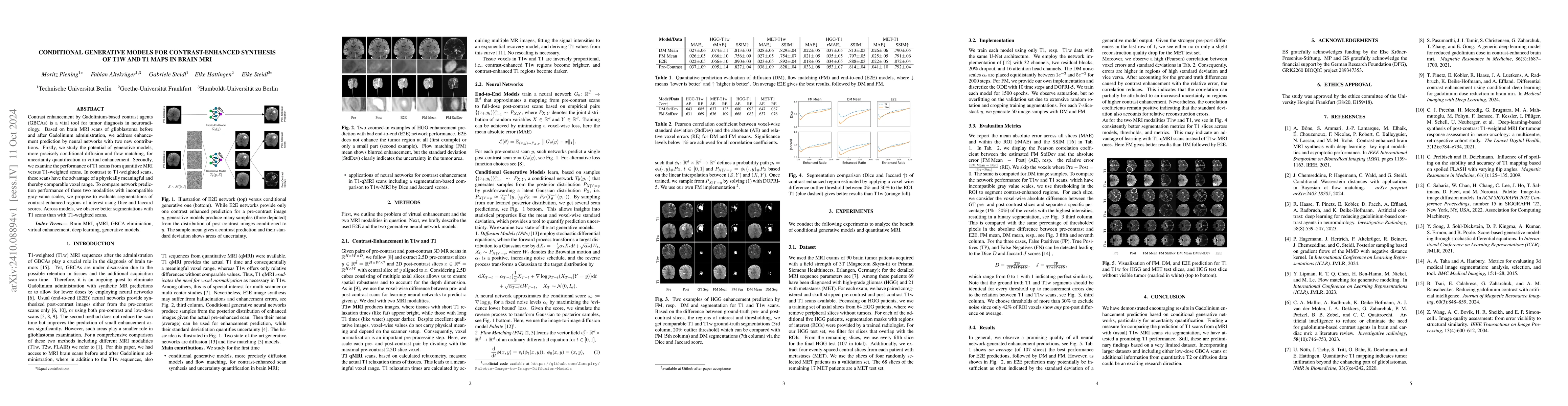
Contrast enhancement by Gadolinium-based contrast agents (GBCAs) is a vital tool for tumor diagnosis in neuroradiology. Based on brain MRI scans of glioblastoma before and after Gadolinium administration, we address enhancement prediction by neural networks with two new contributions. Firstly, we study the potential of generative models, more precisely conditional diffusion and flow matching, for uncertainty quantification in virtual enhancement. Secondly, we examine the performance of T1 scans from quantitive MRI versus T1-weighted scans. In contrast to T1-weighted scans, these scans have the advantage of a physically meaningful and thereby comparable voxel range. To compare network prediction performance of these two modalities with incompatible gray-value scales, we propose to evaluate segmentations of contrast-enhanced regions of interest using Dice and Jaccard scores. Across models, we observe better segmentations with T1 scans than with T1-weighted scans.
AI Key Findings
Get AI-generated insights about this paper's methodology, results, and significance.
Paper Details
PDF Preview
Citation Network
Current paper (gray), citations (green), references (blue)
Display is limited for performance on very large graphs.
Similar Papers
Found 4 papersCAVM: Conditional Autoregressive Vision Model for Contrast-Enhanced Brain Tumor MRI Synthesis
Lujun Gui, Chuyang Ye, Tianyi Yan
Trustworthy Contrast-enhanced Brain MRI Synthesis
Yuxin Li, Xin Gao, Jiyao Liu et al.
T1-contrast Enhanced MRI Generation from Multi-parametric MRI for Glioma Patients with Latent Tumor Conditioning
Xiaofeng Yang, Mojtaba Safari, Zach Eidex et al.
No citations found for this paper.
Comments (0)