Authors
Publication
Metrics
Quick Actions
Quick Answers
What methodology did the authors use?
The research involved designing and fabricating a gamma photon spectrometer using 180nm CMOS technology, integrating configurable analog pixel arrays, SRAM sensitivity calibration units, and decay time processing units to achieve real-time detection of residual cancer cells tagged with gamma-emitting radioisotopes during robotic guided surgery (RGS). More in Methodology →
What are the key results?
The system demonstrated high linearity with 64Cu activity (R2=0.9942) — Accurate tracking of radioactive decay curves over 107 hours More in Key Results →
Why is this work significant?
This research enables precise, real-time detection of residual cancer cells during RGS, improving surgical outcomes and reducing cancer recurrence risks. The system's performance matches clinical requirements for sentinel lymph node monitoring. More in Significance →
What are the main limitations?
Limited testing with diverse radioisotopes — Potential signal conflicts in high-count environments More in Limitations →
Abstract
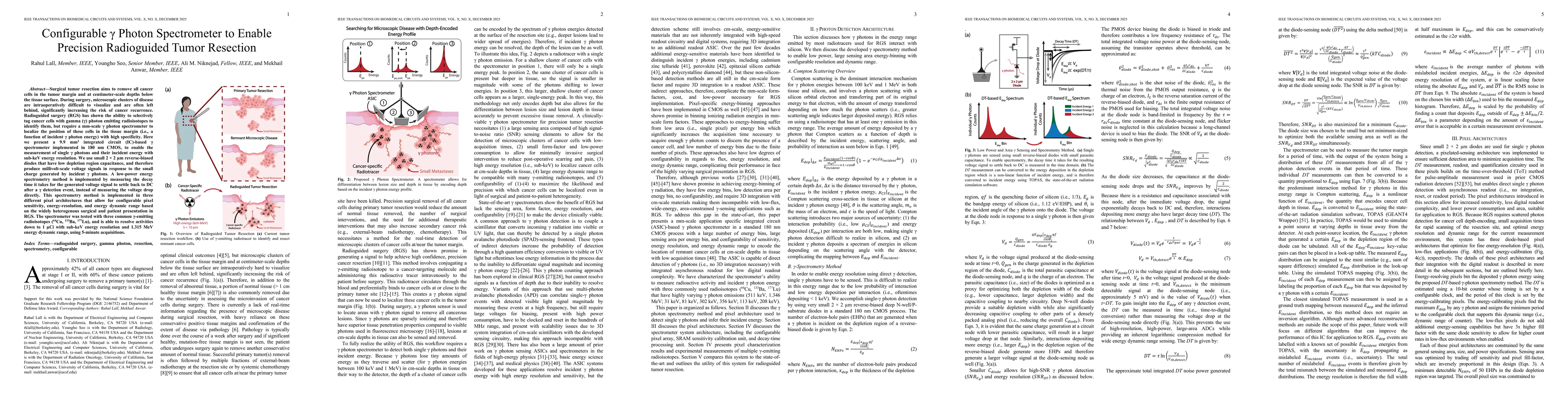
Surgical tumor resection aims to remove all cancer cells in the tumor margin and at centimeter-scale depths below the tissue surface. During surgery, microscopic clusters of disease are intraoperatively difficult to visualize and are often left behind, significantly increasing the risk of cancer recurrence. Radioguided surgery (RGS) has shown the ability to selectively tag cancer cells with gamma (γ) photon emitting radioisotopes to identify them, but require a mm-scale γ photon spectrometer to localize the position of these cells in the tissue margin (i.e., a function of incident γ photon energy) with high specificity. Here we present a 9.9 mm2 integrated circuit (IC)-based γ spectrometer implemented in 180 nm CMOS, to enable the measurement of single γ photons and their incident energy with sub-keV energy resolution. We use small 2 2 um reverse-biased diodes that have low depletion region capacitance, and therefore produce millivolt-scale voltage signals in response to the small charge generated by incident γ photons. A low-power energy spectrometry method is implemented by measuring the decay time it takes for the generated voltage signal to settle back to DC after a γ detection event, instead of measuring the voltage drop directly. This spectrometry method is implemented in three different pixel architectures that allow for configurable pixel sensitivity, energy-resolution, and energy dynamic range based on the widely heterogenous surgical and patient presentation in RGS. The spectrometer was tested with three common γ-emitting radioisotopes (64Cu, 133Ba, 177Lu), and is able to resolve activities down to 1 uCi with sub-keV energy resolution and 1.315 MeV energy dynamic range, using 5-minute acquisitions.
AI Key Findings
Generated Dec 17, 2025
Methodology — What approach did the authors take?
The research involved designing and fabricating a gamma photon spectrometer using 180nm CMOS technology, integrating configurable analog pixel arrays, SRAM sensitivity calibration units, and decay time processing units to achieve real-time detection of residual cancer cells tagged with gamma-emitting radioisotopes during robotic guided surgery (RGS).
Key Results — What are the main findings?
- The system demonstrated high linearity with 64Cu activity (R2=0.9942)
- Accurate tracking of radioactive decay curves over 107 hours
- Sub-keV energy resolution (approx 0.2 keV) for 133Ba and 177Lu
- Depth resolution <1mm for gamma energies 100keV-1MeV
Significance — Why does this research matter?
This research enables precise, real-time detection of residual cancer cells during RGS, improving surgical outcomes and reducing cancer recurrence risks. The system's performance matches clinical requirements for sentinel lymph node monitoring.
Technical Contribution — What is the technical contribution?
Development of a configurable analog pixel array with SRAM-based sensitivity calibration and decay time processing for gamma photon detection in RGS
Novelty — What is new about this work?
Integration of configurable energy resolution and dynamic range with real-time decay time measurement for gamma photon spectroscopy in a compact CMOS platform
Limitations — What are the limitations of this study?
- Limited testing with diverse radioisotopes
- Potential signal conflicts in high-count environments
- Assumptions about silicon interconnects affecting simulation accuracy
Future Work — What did the authors propose for future work?
- Expand testing with additional radioisotopes
- Improve conflict resolution in high-count scenarios
- Refine simulations to include silicon interconnect effects
Paper Details
How to Cite This Paper
@article{niknejad2025configurable,
title = {Configurable γ Photon Spectrometer to Enable Precision Radioguided Tumor Resection},
author = {Niknejad, Ali M. and Anwar, Mekhail and Lall, Rahul and others},
year = {2025},
eprint = {2512.14667},
archivePrefix = {arXiv},
primaryClass = {eess.IV},
doi = {10.1109/TBCAS.2025.3625580},
}
Niknejad, A., Anwar, M., Lall, R., & Seo, Y. (2025). Configurable γ Photon Spectrometer to Enable Precision Radioguided Tumor Resection. arXiv. https://doi.org/10.1109/TBCAS.2025.3625580
Niknejad, Ali M., et al. "Configurable γ Photon Spectrometer to Enable Precision Radioguided Tumor Resection." arXiv, 2025, doi.org/10.1109/TBCAS.2025.3625580.
PDF Preview
Similar Papers
Found 4 papersConfigurable γ Photon Spectrometer to Enable Precision Radioguided Tumor Resection.
Seo, Youngho, Lall, Rahul, Niknejad, Ali M et al.
Advanced Imaging and Localization Techniques in Brain Tumor Resection: A Review for Precision Tumor Removal.
Wei, Jin, Ming, Dong, Liu, Xiuyun et al.
An Inexpensive, Configurable Two-Tone Electron Spin Resonance Spectrometer
Charles A. Collett, Sofia M. Davvetas, Abdulelah Alsuhaymi et al.
Performance of an LYSO-Based Active Converter for a Photon Pair-Spectrometer aiming for 52.8 MeV photon detection in Future $μ^+ \to e^+ γ$ Search Experiments
Sei Ban, Lukas Gerritzen, Fumihito Ikeda et al.
Comments (0)