Confocal Raman microscopy inside sessile multicomponent droplets
Publication
Metrics
Paper Preview
Abstract
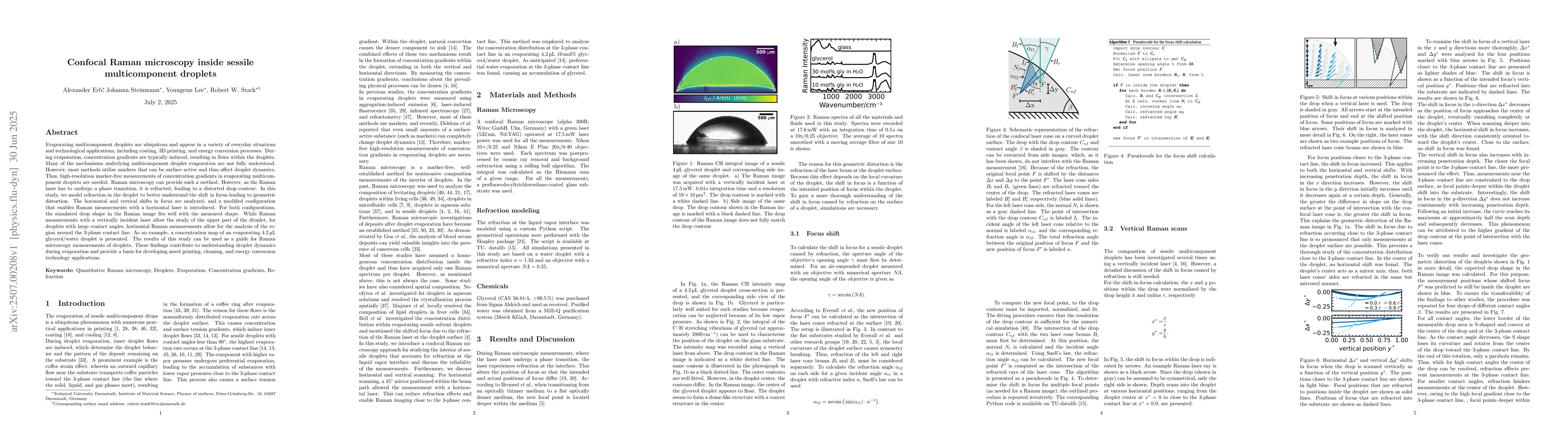
Evaporating multicomponent droplets are ubiquitous and appear in a variety of everyday situations and technological applications, including coating, 3D printing, and energy conversion processes. During evaporation, concentration gradients are typically induced, resulting in flows within the droplets. Many of the mechanisms underlying multicomponent droplet evaporation are not fully understood. However, most methods utilize markers that can be surface active and thus affect droplet dynamics. Thus, high-resolution marker-free measurements of concentration gradients in evaporating multicomponent droplets are needed. Raman microscopy can provide such a method. However, as the Raman laser has to undergo a phase transition, it is refracted, leading to a distorted drop contour. In this study, we model refraction in the droplet to better understand the shift in focus leading to geometric distortion. The horizontal and vertical shifts in focus are analyzed, and a modified configuration that enables Raman measurements with a horizontal laser is introduced. For both configurations, the simulated drop shape in the Raman image fits well with the measured shape. While Raman measurements with a vertically incident laser allow the study of the upper part of the droplet, for droplets with large contact angles, horizontal Raman measurements allow for the analysis of the region around the 3-phase contact line. As an example, a concentration map of an evaporating 4.2 $\mu$L glycerol/water droplet is presented. The results of this study can be used as a guide for Raman microscopy measurements of droplets. These findings contribute to understanding droplet dynamics during evaporation and provide a basis for developing novel printing, cleaning, and energy conversion technology applications.
OpenAlex
- Citations in OpenAlex
- 0
- Publication year
- 2025
- Access status
- Open access
Source: OpenAlex · Snapshot
AI Key Findings
Get AI-generated insights about this paper's methodology, results, significance, and more — seven facets brought into focus.
Impact
Paper Details
Authors
PDF Preview
Citation Network
Current paper (gray), citations (green), references (blue)
Display is limited for performance on very large graphs.
Discussion 0