Continuous max-flow augmentation of self-supervised few-shot learning on SPECT left ventricles
Publication
Metrics
AI Quick Summary
This paper proposes a self-supervised few-shot learning approach combining Continuous Max-Flow augmentation with prior shape information to improve the automatic segmentation of left ventricles in SPECT images, achieving a 5-10% performance increase over existing methods. The method aims to provide a cost-effective solution for reliable myocardial function evaluation.
Paper Preview
Abstract
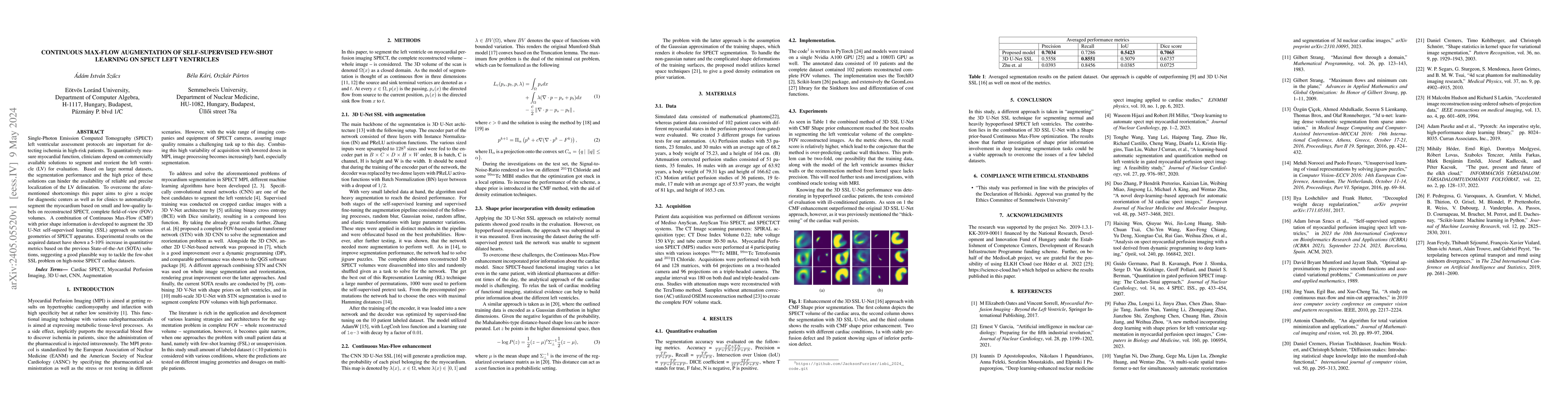
Single-Photon Emission Computed Tomography (SPECT) left ventricular assessment protocols are important for detecting ischemia in high-risk patients. To quantitatively measure myocardial function, clinicians depend on commercially available solutions to segment and reorient the left ventricle (LV) for evaluation. Based on large normal datasets, the segmentation performance and the high price of these solutions can hinder the availability of reliable and precise localization of the LV delineation. To overcome the aforementioned shortcomings this paper aims to give a recipe for diagnostic centers as well as for clinics to automatically segment the myocardium based on small and low-quality labels on reconstructed SPECT, complete field-of-view (FOV) volumes. A combination of Continuous Max-Flow (CMF) with prior shape information is developed to augment the 3D U-Net self-supervised learning (SSL) approach on various geometries of SPECT apparatus. Experimental results on the acquired dataset have shown a 5-10\% increase in quantitative metrics based on the previous State-of-the-Art (SOTA) solutions, suggesting a good plausible way to tackle the few-shot SSL problem on high-noise SPECT cardiac datasets.
AI Key Findings
Get AI-generated insights about this paper's methodology, results, significance, and more — seven facets brought into focus.
Impact
Paper Details
Authors
PDF Preview
Key Terms
Citation Network
Current paper (gray), citations (green), references (blue)
Display is limited for performance on very large graphs.
Discussion 0