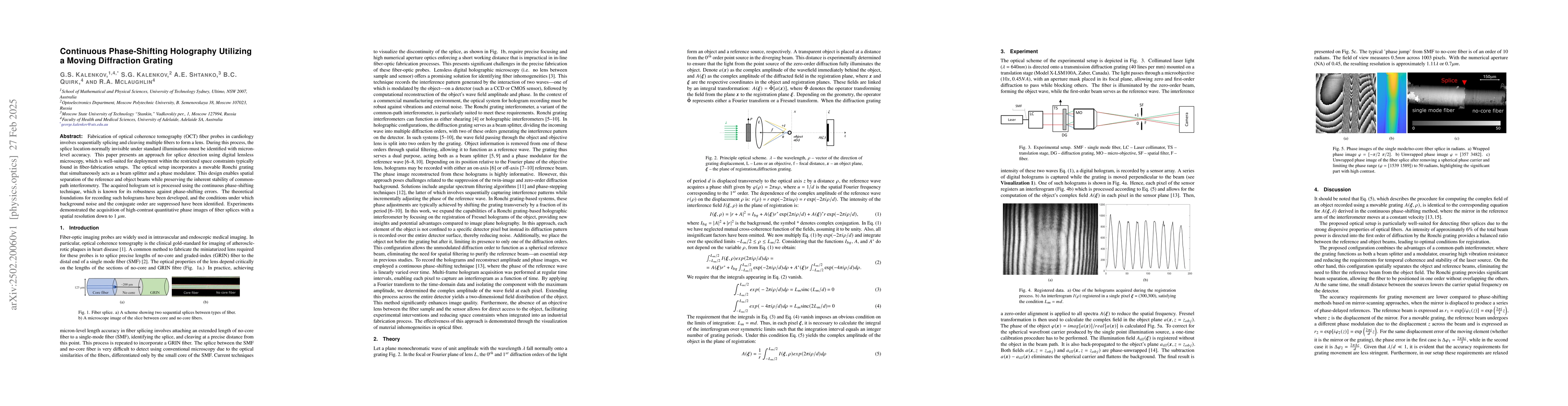
Fabrication of optical coherence tomography (OCT) fiber probes in cardiology
involves sequentially splicing and cleaving multiple fibers to form a lens.
During this process, the splice location-normally invisible under standard
illumination-must be identified with micron-level accuracy. This paper presents
an approach for splice detection using digital lensless microscopy, which is
well-suited for deployment within the restricted space constraints typically
found in fiber-fabrication setups. The optical setup incorporates a movable
Ronchi grating that simultaneously acts as a beam splitter and a phase
modulator. This design enables spatial separation of the reference and object
beams while preserving the inherent stability of common-path interferometry.
The acquired hologram set is processed using the continuous phase-shifting
technique, which is known for its robustness against phase-shifting errors. The
theoretical foundations for recording such holograms have been developed, and
the conditions under which background noise and the conjugate order are
suppressed have been identified. Experiments demonstrated the acquisition of
high-contrast quantitative phase images of fiber splices with a spatial
resolution down to 1 $\mu m$.
Discussion 0