Publication
Metrics
AI Quick Summary
This paper proposes a contrastive learning framework for mitochondria segmentation in electron microscopy images, utilizing a point sampling strategy and a pixel-wise label-based contrastive loss to improve feature representation. The method achieves better or on par segmentation results compared to state-of-the-art techniques on MitoEM and FIB-SEM datasets.
Paper Preview
Abstract
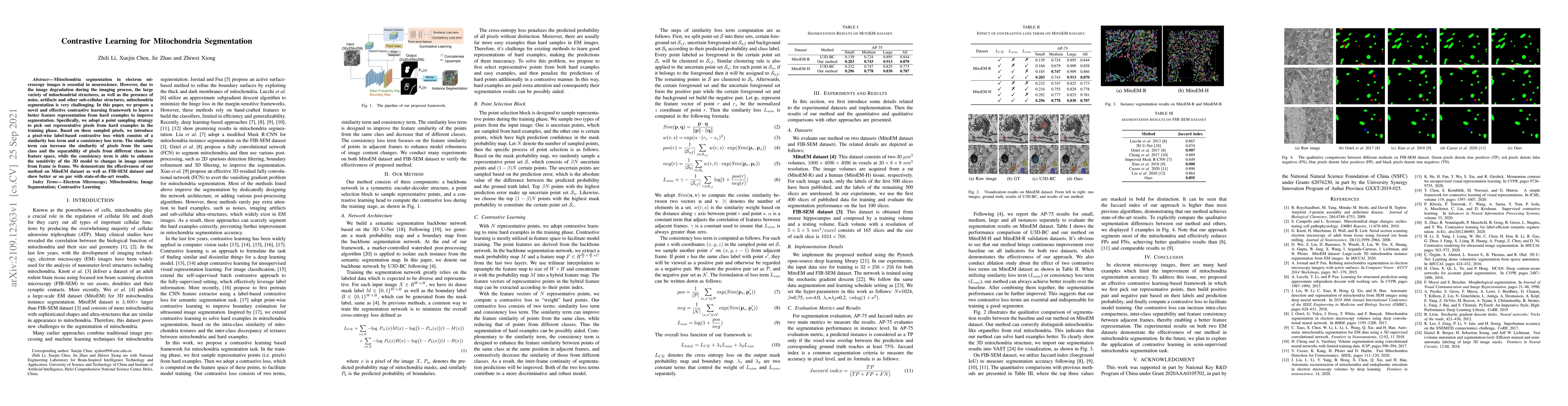
Mitochondria segmentation in electron microscopy images is essential in neuroscience. However, due to the image degradation during the imaging process, the large variety of mitochondrial structures, as well as the presence of noise, artifacts and other sub-cellular structures, mitochondria segmentation is very challenging. In this paper, we propose a novel and effective contrastive learning framework to learn a better feature representation from hard examples to improve segmentation. Specifically, we adopt a point sampling strategy to pick out representative pixels from hard examples in the training phase. Based on these sampled pixels, we introduce a pixel-wise label-based contrastive loss which consists of a similarity loss term and a consistency loss term. The similarity term can increase the similarity of pixels from the same class and the separability of pixels from different classes in feature space, while the consistency term is able to enhance the sensitivity of the 3D model to changes in image content from frame to frame. We demonstrate the effectiveness of our method on MitoEM dataset as well as FIB-SEM dataset and show better or on par with state-of-the-art results.
AI Key Findings — Failed
Key findings generation failed. Failed to start generation process
Impact
Paper Details
Authors
PDF Preview
Key Terms
Citation Network
Current paper (gray), citations (green), references (blue)
Display is limited for performance on very large graphs.
Discussion 0