This research paper introduces a one-shot method for object localization in 3D medical images that doesn't require annotated data, instead relying on relative position prediction to locate organs and landmarks.
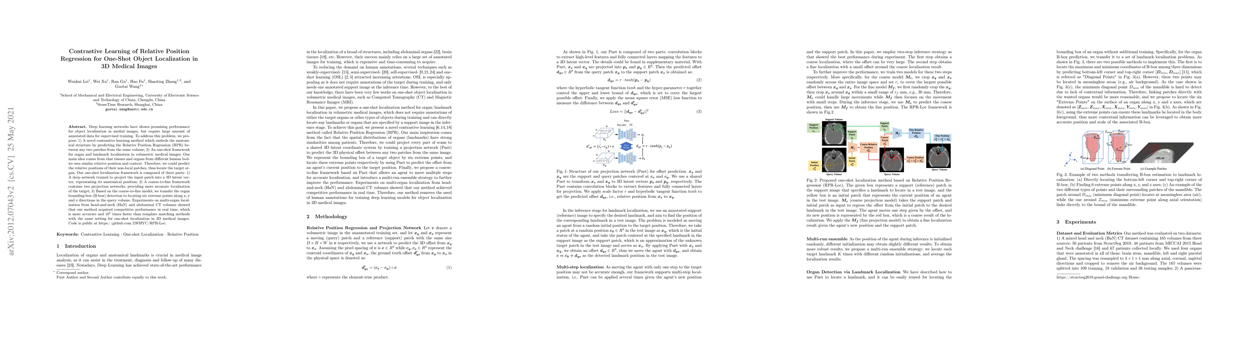
Deep learning networks have shown promising performance for accurate object localization in medial images, but require large amount of annotated data for supervised training, which is expensive and expertise burdensome. To address this problem, we present a one-shot framework for organ and landmark localization in volumetric medical images, which does not need any annotation during the training stage and could be employed to locate any landmarks or organs in test images given a support (reference) image during the inference stage. Our main idea comes from that tissues and organs from different human bodies have a similar relative position and context. Therefore, we could predict the relative positions of their non-local patches, thus locate the target organ. Our framework is composed of three parts: (1) A projection network trained to predict the 3D offset between any two patches from the same volume, where human annotations are not required. In the inference stage, it takes one given landmark in a reference image as a support patch and predicts the offset from a random patch to the corresponding landmark in the test (query) volume. (2) A coarse-to-fine framework contains two projection networks, providing more accurate localization of the target. (3) Based on the coarse-to-fine model, we transfer the organ boundingbox (B-box) detection to locating six extreme points along x, y and z directions in the query volume. Experiments on multi-organ localization from head-and-neck (HaN) CT volumes showed that our method acquired competitive performance in real time, which is more accurate and 10^5 times faster than template matching methods with the same setting. Code is available: https://github.com/LWHYC/RPR-Loc.
Get AI-generated insights about this paper's methodology, results, and significance.
Current paper (gray), citations (green), references (blue)
Yilei Shi, Xiao Xiang Zhu, Chunlei Li et al.
Xiang Li, Jiawei Zhou, Yi Cao et al.
Title | Authors | Year | Actions |
---|---|---|---|
BodyGPS: Anatomical Positioning System
|
H. Yerebakan, Kritika Iyer, Xueqi Guo, Y. Shinagawa, G. H. Valadez
|
2025
|
View Paper |
Learning Consistent Semantic Representation for Chest X-ray via Anatomical Localization in Self-Supervised Pre-Training
|
Surong Chu, Xueting Ren, Guo-hua Ji, Juanjuan Zhao, Jinwei Shi, Yangyang Wei, Bo Pei, Yan Qiang
|
2025
|
View Paper |
MedLSAM: Localize and segment anything model for 3D CT images
|
Wenhui Lei, Wei Xu, Kang Li, Xiaofan Zhang, Shaoting Zhang
|
2024
|
View Paper |
Anatomical Positional Embeddings
|
M. Goncharov, Valentin Samokhin, Eugenia Soboleva, Roman Sokolov, B. Shirokikh, Mikhail Belyaev, Anvar Kurmukov, Ivan Oseledets
|
2024
|
View Paper |
Weakly Supervised Lymph Nodes Segmentation Based on Partial Instance Annotations with Pre-trained Dual-branch Network and Pseudo Label Learning
|
Litingyu Wang, Yijie Qu, Xiangde Luo, Wenjun Liao, Shichuan Zhang, Guotai Wang
|
2024
|
View Paper |
ScribSD+: Scribble-supervised medical image segmentation based on simultaneous multi-scale knowledge distillation and class-wise contrastive regularization
|
Yijie Qu, Tao Lu, Shaoting Zhang, Guotai Wang
|
2024
|
View Paper |
Beyond Pixel-Wise Supervision for Medical Image Segmentation: From Traditional Models to Foundation Models
|
Yuyan Shi, Jialu Ma, Jin Yang, Shasha Wang, Yichi Zhang
|
2024
|
View Paper |
DrasCLR: A self-supervised framework of learning disease-related and anatomy-specific representation for 3D lung CT images
|
Ke Yu, Li Sun, Junxiang Chen, Maxwell Reynolds, Tigmanshu Chaudhary, K. Batmanghelich
|
2023
|
View Paper |
Foundational Models Defining a New Era in Vision: A Survey and Outlook
|
Muhammad Awais, Muzammal Naseer, Salman Siddique Khan, R. Anwer, Hisham Cholakkal, M. Shah, Ming Yang, F. Khan
|
2023
|
View Paper |
One-Shot Weakly-Supervised Segmentation in 3D Medical Images
|
Wenhui Lei, Qi Su, Tianyu Jiang, Ran Gu, Na Wang, Xinglong Liu, Guotai Wang, Xiaofan Zhang, Shaoting Zhang
|
2023
|
View Paper |
Position Regression for Unsupervised Anomaly Detection
|
Florentin Bieder, Julia Wolleb, Robin Sandkühler, P. Cattin
|
2023
|
View Paper |
Researches advanced in application of medical image analysis based on deep learning
|
Xichen Hu, Z. Guo, Sheng Yang, Kaiyuan Zheng
|
2022
|
View Paper |
CDDSA: Contrastive Domain Disentanglement and Style Augmentation for Generalizable Medical Image Segmentation
|
Ran Gu, Guotai Wang, Jiangshan Lu, Jingyang Zhang, Wenhui Lei, Yinan Chen, Wenjun Liao, Shichuan Zhang, Kang Li, Dimitris N. Metaxas, Shaoting Zhang
|
2022
|
View Paper |
Information-guided pixel augmentation for pixel-wise contrastive learning
|
Quan Quan, Qingsong Yao, Jun Li, S.kevin Zhou
|
2022
|
View Paper |
Adaptive Contrastive Learning with Dynamic Correlation for Multi-Phase Organ Segmentation
|
Ho Hin Lee, Yucheng Tang, Han Liu, Yubo Fan, L. Cai, Qi Yang, Xin Yu, Shunxing Bao, Yuankai Huo, Bennett A. Landman
|
2022
|
View Paper |
TractoSCR: a novel supervised contrastive regression framework for prediction of neurocognitive measures using multi-site harmonized diffusion MRI tractography
|
Tengfei Xue, Fan Zhang, Leo R. Zekelman, Chaoyi Zhang, Yuqian Chen, S. Cetin-Karayumak, Steve Pieper, W. Wells, Y. Rathi, N. Makris, Weidong Cai, L. O’Donnell
|
2022
|
View Paper |
Dissecting Self-Supervised Learning Methods for Surgical Computer Vision
|
Sanat Ramesh, V. Srivastav, Deepak Alapatt, Tong Yu, Aditya Murali, Luca Sestini, C. Nwoye, Idris Hamoud, A. Fleurentin, Georgios Exarchakis, A. Karargyris, N. Padoy
|
2022
|
View Paper |
Deep learning of birth-related infant clavicle fractures: a potential virtual consultant for fracture dating
|
Andy Tsai, P. Grant, S. Warfield, Yangming Ou, P. Kleinman
|
2022
|
View Paper |
Relative distance matters for one-shot landmark detection
|
Qingsong Yao, Jianji Wang, Yihua Sun, Quan Quan, Heqin Zhu, S. K. Zhou
|
2022
|
View Paper |
Machine learning to identify distal tibial classic metaphyseal lesions of infant abuse: a pilot study
|
Andy Tsai, P. Kleinman
|
2022
|
View Paper |
Intra- and Inter-Slice Contrastive Learning for Point Supervised OCT Fluid Segmentation
|
Xingxin He, Leyuan Fang, Mingkui Tan, Xiangdong Chen
|
2022
|
View Paper |
Which images to label for few-shot medical landmark detection?
|
Quan Quan, Qingsong Yao, Jun Li, S. K. Zhou
|
2021
|
View Paper |
One-shot Weakly-Supervised Segmentation in Medical Images
|
Wenhui Lei, Q. Su, Ran Gu, Na Wang, Xinglong Liu, Guotai Wang, Xiaofan Zhang, Shaoting Zhang
|
2021
|
View Paper |
Unsupervised Local Discrimination for Medical Images
|
Huai Chen, Renzhen Wang, Jieyu Li, Qing Peng, Deyu Meng, Lisheng Wang
|
2021
|
View Paper |
MedLSAM: Localize and Segment Anything Model for 3D Medical Images
|
Wenhui Lei, Xu Wei, Xiaofan Zhang, Kang Li, Shaoting Zhang
|
2023
|
View Paper |
A Novel Supervised Contrastive Regression Framework for Prediction of Neurocognitive Measures Using Multi-Site Harmonized Diffusion MRI Tractography
|
Tengfei Xue, Fan Zhang, Leo R. Zekelman, Chaoyi Zhang, Yuqian Chen, Suheyla Cetin Karayumak, Steve Pieper, William M. Wells, Y. Rathi, N. Makris, Weidong Cai, L. O’Donnell
|
2022
|
View Paper |
Medical Image Analysis
|
Wenhui Lei, Wei Xu, Kang Li, Xiaofan Zhang, Shaoting Zhang
|
View Paper | |
Automatic Segmentation of Abdominal Organs with 3D-UNet
|
Wenhui Lei
|
View Paper |
Comments (0)