Publication
Metrics
AI Quick Summary
This paper proposes a contrastive learning approach to automatically segment cytoarchitectonic areas in histological human brain sections, aiming to map the entire brain. The method encodes microscopic image patches into robust features for area classification, demonstrating improved performance over models trained from scratch or on auxiliary tasks, and showing anatomically meaningful groupings in feature space.
Paper Preview
Abstract
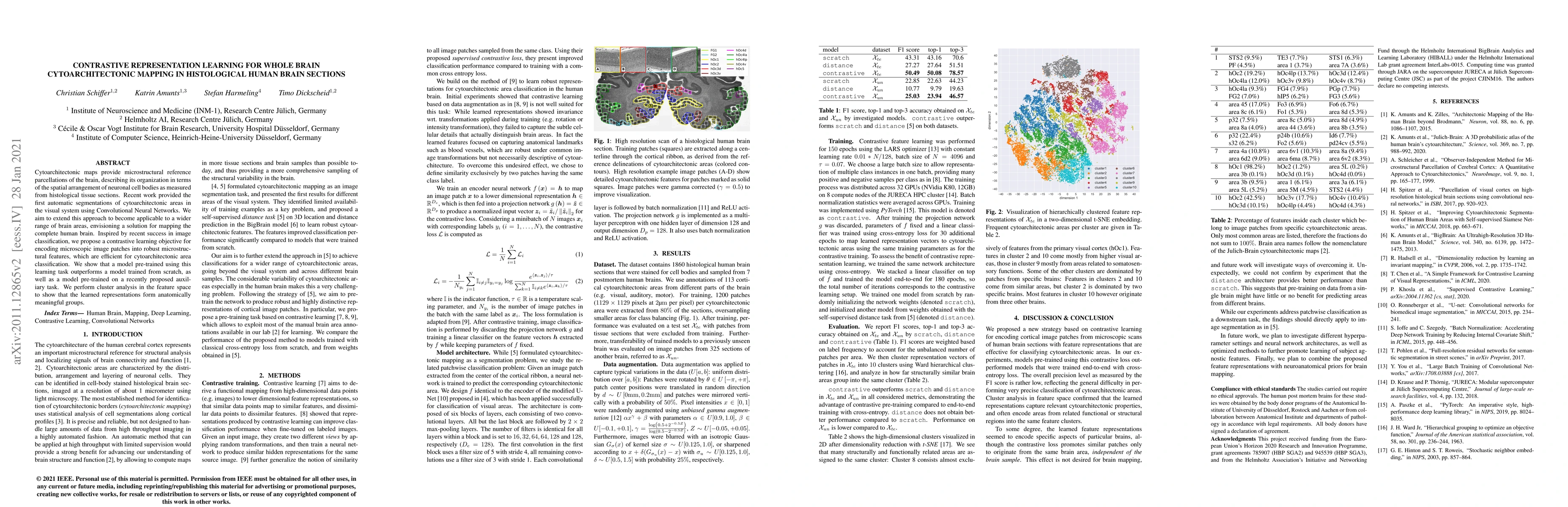
Cytoarchitectonic maps provide microstructural reference parcellations of the brain, describing its organization in terms of the spatial arrangement of neuronal cell bodies as measured from histological tissue sections. Recent work provided the first automatic segmentations of cytoarchitectonic areas in the visual system using Convolutional Neural Networks. We aim to extend this approach to become applicable to a wider range of brain areas, envisioning a solution for mapping the complete human brain. Inspired by recent success in image classification, we propose a contrastive learning objective for encoding microscopic image patches into robust microstructural features, which are efficient for cytoarchitectonic area classification. We show that a model pre-trained using this learning task outperforms a model trained from scratch, as well as a model pre-trained on a recently proposed auxiliary task. We perform cluster analysis in the feature space to show that the learned representations form anatomically meaningful groups.
AI Key Findings
Get AI-generated insights about this paper's methodology, results, significance, and more — seven facets brought into focus.
Impact
Paper Details
PDF Preview
Key Terms
Citation Network
Current paper (gray), citations (green), references (blue)
Display is limited for performance on very large graphs.
Discussion 0