Convolutional nets for reconstructing neural circuits from brain images acquired by serial section electron microscopy
Publication
Metrics
AI Quick Summary
This paper explores the application of convolutional neural networks (CNNs) to automate the reconstruction of neural circuits from brain images obtained via serial section electron microscopy, addressing challenges in image defect handling and expanding tasks beyond neuronal boundary detection. Computational systems are being developed to manage the vast data volumes inherent in this imaging technique.
Paper Preview
Abstract
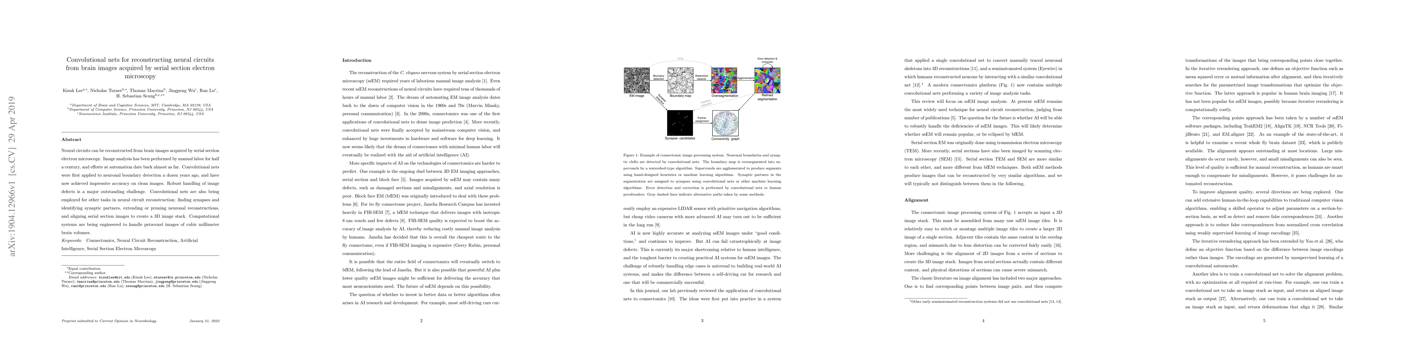
Neural circuits can be reconstructed from brain images acquired by serial section electron microscopy. Image analysis has been performed by manual labor for half a century, and efforts at automation date back almost as far. Convolutional nets were first applied to neuronal boundary detection a dozen years ago, and have now achieved impressive accuracy on clean images. Robust handling of image defects is a major outstanding challenge. Convolutional nets are also being employed for other tasks in neural circuit reconstruction: finding synapses and identifying synaptic partners, extending or pruning neuronal reconstructions, and aligning serial section images to create a 3D image stack. Computational systems are being engineered to handle petavoxel images of cubic millimeter brain volumes.
AI Key Findings
Get AI-generated insights about this paper's methodology, results, significance, and more — seven facets brought into focus.
Impact
Paper Details
PDF Preview
Key Terms
Citation Network
Current paper (gray), citations (green), references (blue)
Display is limited for performance on very large graphs.
Discussion 0