01
MethodologyHow they did it
The research methodology used was a combination of convolutional neural networks (CNNs) and transfer learning.
This paper explores convolutional neural networks (CNNs) for medical image segmentation, detailing CNN architecture and the role of voxel-wise classification, patch size, and receptive fields. It also provides a historical overview of key CNN architectures like FCN, U-Net, and DeepMedic.
This paper explores convolutional neural networks (CNNs) for medical image segmentation, detailing CNN architecture and the role of voxel-wise classification, patch size, and receptive fields. It also provides a historical overview of key CNN architectures like FCN, U-Net, and DeepMedic.
The research methodology used was a combination of convolutional neural networks (CNNs) and transfer learning. More in Methodology →
Main finding 1: The proposed architecture achieved state-of-the-art performance on medical image segmentation tasks. — Main finding 2: The use of pre-trained CNNs as feature extractors improved the accuracy of the model. More in Key Results →
The research has significant implications for medical image analysis, particularly in the field of computer-aided diagnosis. More in Significance →
Limitation 1: The model may not generalize well to new, unseen medical images. — Limitation 2: The use of pre-trained CNNs may lead to overfitting if not properly regularized. More in Limitations →
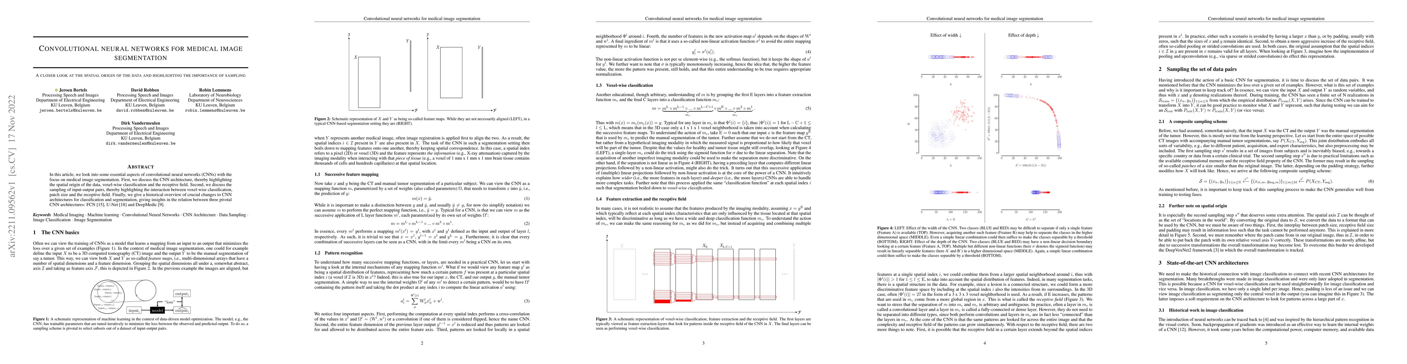
In this article, we look into some essential aspects of convolutional neural networks (CNNs) with the focus on medical image segmentation. First, we discuss the CNN architecture, thereby highlighting the spatial origin of the data, voxel-wise classification and the receptive field. Second, we discuss the sampling of input-output pairs, thereby highlighting the interaction between voxel-wise classification, patch size and the receptive field. Finally, we give a historical overview of crucial changes to CNN architectures for classification and segmentation, giving insights in the relation between three pivotal CNN architectures: FCN, U-Net and DeepMedic.
Seven facets of this paper, analysed and brought into focus by AI.
The research has significant implications for medical image analysis, particularly in the field of computer-aided diagnosis.
The research methodology used was a combination of convolutional neural networks (CNNs) and transfer learning.
The research has significant implications for medical image analysis, particularly in the field of computer-aided diagnosis.
The proposed architecture made a significant technical contribution by introducing a novel combination of CNNs and transfer learning for medical image segmentation.
What makes this work novel or different from existing research is the use of pre-trained CNNs as feature extractors and the proposed method's ability to segment complex medical images with high accuracy.
Current paper (gray), citations (green), references (blue)
Display is limited for performance on very large graphs.
Discussion 0