Core language brain network for fMRI-language task used in clinical applications
Publication
Metrics
AI Quick Summary
Researchers mapped the brain network for language tasks in healthy individuals, identifying a common architecture involving Broca's area, Wernicke's area, and others. The study's findings provide insights for clinical applications and highlight important connections that should be preserved through brain surgery.
Paper Preview
Abstract
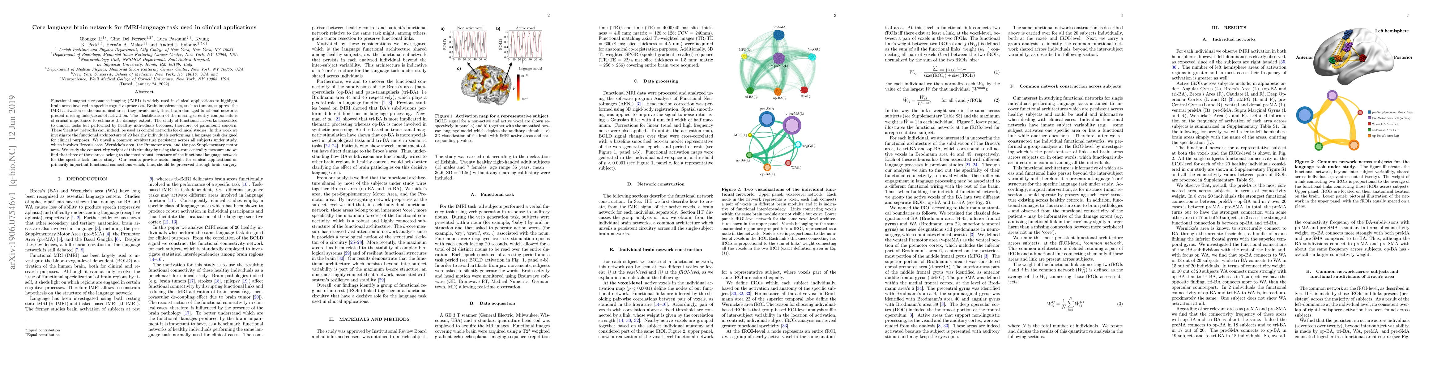
Functional magnetic resonance imaging (fMRI) is widely used in clinical applications to highlight brain areas involved in specific cognitive processes. Brain impairments, such as tumors, suppress the fMRI activation of the anatomical areas they invade and, thus, brain-damaged functional networks present missing links/areas of activation. The identification of the missing circuitry components is of crucial importance to estimate the damage extent. The study of functional networks associated to clinical tasks but performed by healthy individuals becomes, therefore, of paramount concern. These `healthy' networks can, indeed, be used as control networks for clinical studies. In this work we investigate the functional architecture of 20 healthy individuals performing a language task designed for clinical purposes. We unveil a common architecture persistent across all subjects under study, which involves Broca's area, Wernicke's area, the Premotor area, and the pre-Supplementary motor area. We study the connectivity weight of this circuitry by using the k-core centrality measure and we find that three of these areas belong to the most robust structure of the functional language network for the specific task under study. Our results provide useful insight for clinical applications on primarily important functional connections which, thus, should be preserved through brain surgery.
AI Key Findings
Get AI-generated insights about this paper's methodology, results, significance, and more — seven facets brought into focus.
Impact
Paper Details
PDF Preview
Key Terms
Citation Network
Current paper (gray), citations (green), references (blue)
Display is limited for performance on very large graphs.
Discussion 0