Publication
Metrics
AI Quick Summary
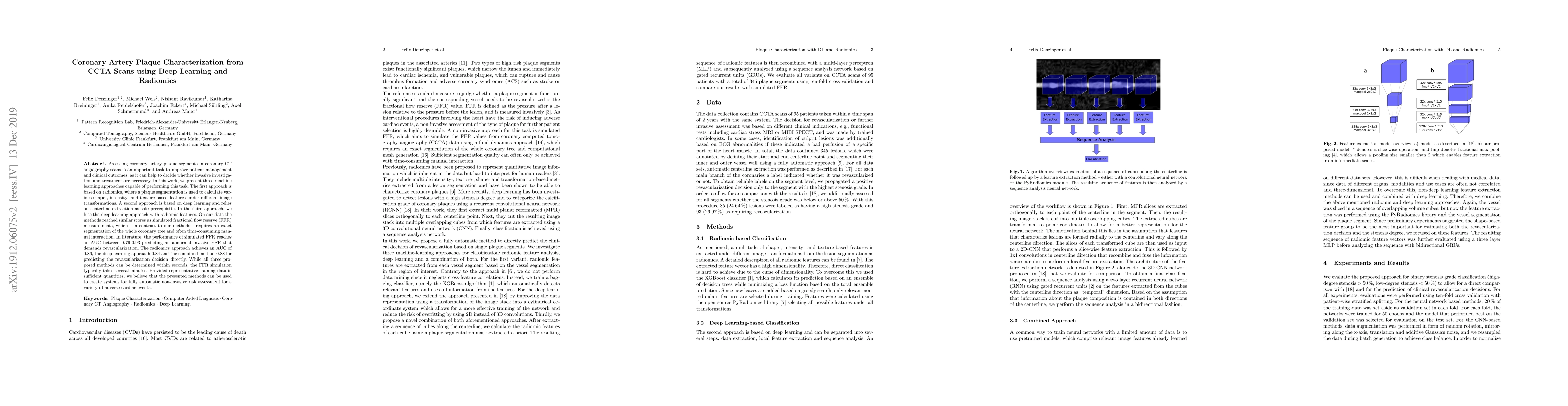
This paper presents three machine learning approaches for characterizing coronary artery plaques from CT angiography scans: a radiomics-based method, a deep learning approach, and a fusion of both. The methods achieve high AUC scores (0.86-0.88) for predicting revascularization needs, outperforming the time-consuming simulated fractional flow reserve method.
Paper Preview
Abstract
Assessing coronary artery plaque segments in coronary CT angiography scans is an important task to improve patient management and clinical outcomes, as it can help to decide whether invasive investigation and treatment are necessary. In this work, we present three machine learning approaches capable of performing this task. The first approach is based on radiomics, where a plaque segmentation is used to calculate various shape-, intensity- and texture-based features under different image transformations. A second approach is based on deep learning and relies on centerline extraction as sole prerequisite. In the third approach, we fuse the deep learning approach with radiomic features. On our data the methods reached similar scores as simulated fractional flow reserve (FFR) measurements, which - in contrast to our methods - requires an exact segmentation of the whole coronary tree and often time-consuming manual interaction. In literature, the performance of simulated FFR reaches an AUC between 0.79-0.93 predicting an abnormal invasive FFR that demands revascularization. The radiomics approach achieves an AUC of 0.86, the deep learning approach 0.84 and the combined method 0.88 for predicting the revascularization decision directly. While all three proposed methods can be determined within seconds, the FFR simulation typically takes several minutes. Provided representative training data in sufficient quantities, we believe that the presented methods can be used to create systems for fully automatic non-invasive risk assessment for a variety of adverse cardiac events.
AI Key Findings
Get AI-generated insights about this paper's methodology, results, significance, and more — seven facets brought into focus.
Impact
Paper Details
PDF Preview
Key Terms
Citation Network
Current paper (gray), citations (green), references (blue)
Display is limited for performance on very large graphs.
Discussion 0