ArxivLens

Quick Summary:

This paper presents DeepCap, a deep learning model based on capsule networks for segmenting coronary arteries from intravascular optical coherence tomography (IVOCT) images, achieving high segmentation quality and efficiency with a small memory footprint and fast inference times. The model is shown to be robust and comparable to state-of-the-art methods, offering a valuable tool for clinicians.

DOI: 10.1016/j.artmed.2021.102072
arXiv Id: 2003.06080
Comments: This version has been accepted in Artificial Intelligence in Medicine. Main paper: 28 pages, 9 figures, 4 tables. Supplementary Material: 3 pages, 3 figures
Date Published: 2021-04-20
Date Updated: 2021-04-20

Categories:

Key Terms (TF-IDF):

segmentation (0.240)
segmentation (0.239)
labelled (0.207)
expert (0.205)
image processing (0.205)
unbiased (0.200)
expert (0.193)
unbiased (0.179)
automated (0.179)
automated (0.169)
faster (0.167)
dataset (0.165)
image (0.162)
dataset (0.160)
image (0.155)
inference (0.155)
faster (0.153)
processing (0.150)
inference (0.143)
robust (0.138)
License: http://creativecommons.org/licenses/by/4.0/

Summary:

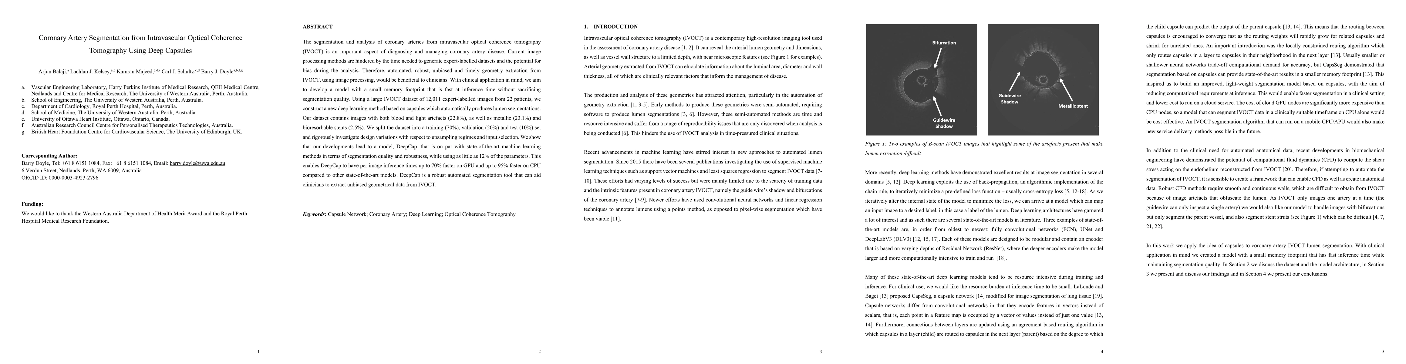
The segmentation and analysis of coronary arteries from intravascular optical coherence tomography (IVOCT) is an important aspect of diagnosing and managing coronary artery disease. Current image processing methods are hindered by the time needed to generate expert-labelled datasets and the potential for bias during the analysis. Therefore, automated, robust, unbiased and timely geometry extraction from IVOCT, using image processing, would be beneficial to clinicians. With clinical application in mind, we aim to develop a model with a small memory footprint that is fast at inference time without sacrificing segmentation quality. Using a large IVOCT dataset of 12,011 expert-labelled images from 22 patients, we construct a new deep learning method based on capsules which automatically produces lumen segmentations. Our dataset contains images with both blood and light artefacts (22.8%), as well as metallic (23.1%) and bioresorbable stents (2.5%). We split the dataset into a training (70%), validation (20%) and test (10%) set and rigorously investigate design variations with respect to upsampling regimes and input selection. We show that our developments lead to a model, DeepCap, that is on par with state-of-the-art machine learning methods in terms of segmentation quality and robustness, while using as little as 12% of the parameters. This enables DeepCap to have per image inference times up to 70% faster on GPU and up to 95% faster on CPU compared to other state-of-the-art models. DeepCap is a robust automated segmentation tool that can aid clinicians to extract unbiased geometrical data from IVOCT.

Citation & Reference Network Visualization

This graph shows how this paper is connected to other research. The current paper is shown in dark gray, papers that cite it are in green, and papers it references are in blue. You can:

Current Paper
Papers citing this work (Citations)
Papers cited by this work (References)

• Hover over any node to see the paper title

• Click a node to view that paper's details

• Drag nodes to rearrange the graph

• Use the zoom controls (bottom right) to zoom in/out

Citations and References

Title Authors Year Actions
Enhancing percutaneous coronary intervention using TriVOCTNet: a multi-task deep learning model for comprehensive intravascular optical coherence tomography analysis.
Yu Shi Lau, Li Kuo Tan, Kok Han Chee, C. Chan, Y. Liew
2025
View Paper
Rapid automated lumen segmentation of coronary optical coherence tomography images followed by 3D reconstruction of coronary arteries
Wei Wu, Merjulah Roby, Akshat Banga, Usama M Oguz, Vinay Kumar Gadamidi, Charu Hasini Vasa, Shijia Zhao, Vineeth S. Dasari, A. Thota, Sartaj Tanweer, Changkye Lee, Ghassan S. Kassab, Y. Chatzizisis
2024
View Paper
Attention-gated 3D CapsNet for robust hippocampal segmentation
Clément Poiret, Antoine Bouyeure, Sandesh Patil, Cécile Boniteau, Edouard Duchesnay, Antoine Grigis, Frédéric Lemaître, Marion Noulhiane
2024
View Paper
Efficient Autonomous Lumen Segmentation in Intravascular Optical Coherence Tomography Images: Unveiling the Potential of Polynomial-Regression Convolutional Neural Network
Yu Shi Lau, Li Kuo Tan, Kok Han Chee, Chow Khuen Chan, Y. Liew
2023
View Paper
Unravelling the role of macrophages in cardiovascular inflammation through imaging: a state-of-the-art review
R. Parry, K. Majeed, F. Pixley, G. Hillis, R. Francis, C. Schultz
2022
View Paper
Automated Coronary Optical Coherence Tomography Feature Extraction with Application to Three-Dimensional Reconstruction
H. Carpenter, M. Ghayesh, A. Zander, Jiawen Li, G. Di Giovanni, P. Psaltis
2022
View Paper
Reduced order models-II
Fausto Dicech, Riccardo Zamolo, Lucia Parussini
View Paper

Similar Papers

Segmentation of Anatomical Layers and Artifacts in Intravascular Polarization Sensitive Optical Coherence Tomography Using Attending Physician and Boundary Cardinality Losses

Polina Golland, Brett E. Bouma, Martin Villiger, Joost Daemen, Mohammad Haft-Javaherian, Kenichiro Otsuka, Peter Libby

Published: 2022-09-15

Morphology-based non-rigid registration of coronary computed tomography and intravascular images through virtual catheter path optimization

Karim Kadry, Michiel Schaap, David Marlevi, Charles Taylor, Abhishek Karmakar, Andreas Schuh, Kersten Peterson, Elazer Edelman, Farhad Nezami

Published: 2023-02-02

Prediction of stent under-expansion in calcified coronary arteries using machine-learning on intravascular optical coherence tomography

Juhwan Lee, David L. Wilson, Ammar Hoori, Sadeer Al-Kindi, Justin N. Kim, Luis A. P. Dallan, Vladislav N. Zimin, Hiram G. Bezerra, Pengfei Dong, Linxia Gu, Gabriel T. R. Pereira, Yazan Gharaibeh, Chaitanya Kolluru, Armando Vergara-Martel, Peshala T. Gamage

Published: 2022-05-24

Comments (0)

This website uses cookies to ensure you get the best experience. Learn more