Cortical Geometry Network and Topology Markers for Parkinson's Disease
Publication
Metrics
AI Quick Summary
A new method uses geometry networks to study brain surface geometry in Parkinson's disease, finding a statistically significant difference between patient and healthy brains.
Paper Preview
Abstract
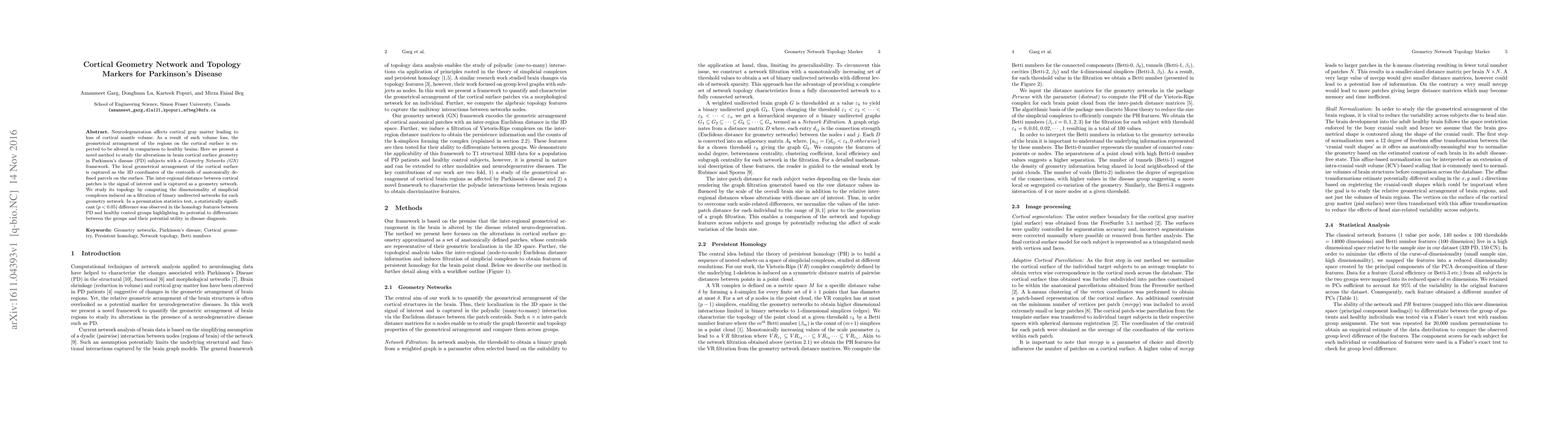
Neurodegeneration affects cortical gray matter leading to loss of cortical mantle volume. As a result of such volume loss, the geometrical arrangement of the regions on the cortical surface is expected to be altered in comparison to healthy brains. Here we present a novel method to study the alterations in brain cortical surface geometry in Parkinson's disease (PD) subjects with a \emph{Geometry Networks (GN)} framework. The local geometrical arrangement of the cortical surface is captured as the 3D coordinates of the centroids of anatomically defined parcels on the surface. The inter-regional distance between cortical patches is the signal of interest and is captured as a geometry network. We study its topology by computing the dimensionality of simplicial complexes induced on a filtration of binary undirected networks for each geometry network. In a permutation statistics test, a statistically significant ($p<0.05$) difference was observed in the homology features between PD and healthy control groups highlighting its potential to differentiate between the groups and their potential utility in disease diagnosis.
AI Key Findings
Get AI-generated insights about this paper's methodology, results, significance, and more — seven facets brought into focus.
Impact
Paper Details
PDF Preview
Key Terms
Citation Network
Current paper (gray), citations (green), references (blue)
Display is limited for performance on very large graphs.
Discussion 0