Publication
Metrics
AI Quick Summary
This research proposes an improved method for COVID-19 detection using CT images, involving slice processing and a modified Xception classifier, which achieved higher validation accuracy and macro F1 score compared to previous methods. The method emphasizes lung areas through slice selection and cropping, followed by input into a resized Xception model.
Paper Preview
Abstract
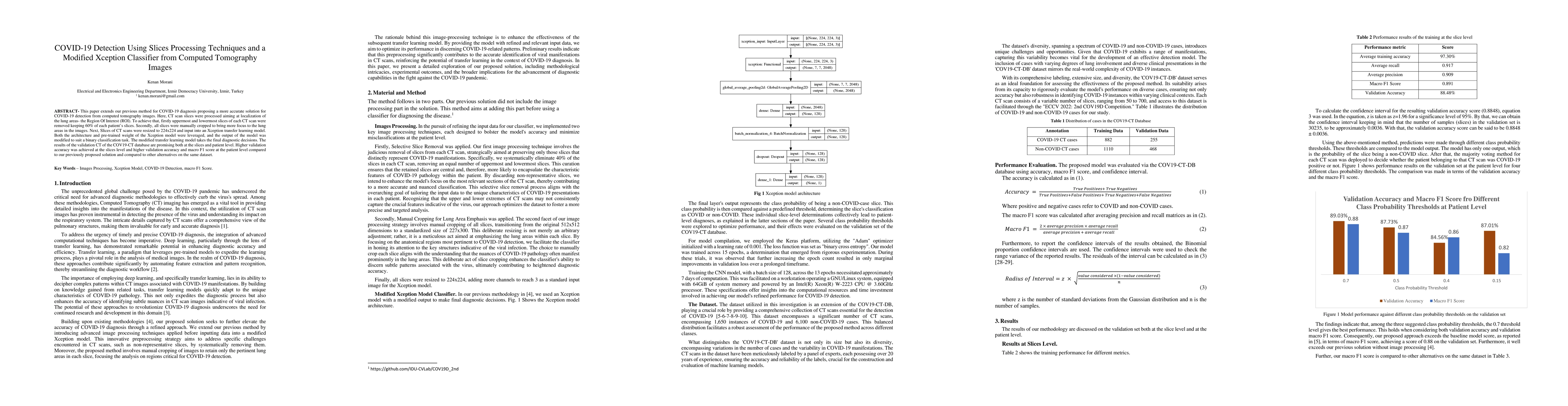
This paper extends our previous method for COVID-19 diagnosis, proposing an enhanced solution for detecting COVID-19 from computed tomography (CT) images. To decrease model misclassifications, two key steps of image processing were employed. Firstly, the uppermost and lowermost slices were removed, preserving sixty percent of each patient's slices. Secondly, all slices underwent manual cropping to emphasize the lung areas. Subsequently, resized CT scans (224 by 224) were input into an Xception transfer learning model. Leveraging Xception's architecture and pre-trained weights, the modified model achieved binary classification. Promising results on the COV19-CT database showcased higher validation accuracy and macro F1 score at both the slice and patient levels compared to our previous solution and alternatives on the same dataset.
AI Key Findings
Get AI-generated insights about this paper's methodology, results, significance, and more — seven facets brought into focus.
Impact
Paper Details
Authors
PDF Preview
Key Terms
Citation Network
Current paper (gray), citations (green), references (blue)
Display is limited for performance on very large graphs.
Discussion 0