Publication
Metrics
AI Quick Summary
This research develops an AI-based classification model for determining COVID-19 severity levels from chest X-ray images, employing pre-processing, image augmentation, and feature extraction using ResNet-50, VGG16, and SVM. The ResNet-50 model achieved superior performance with 95% accuracy, 0.94 recall, 0.92 F1-Score, and 0.91 precision.
Paper Preview
Abstract
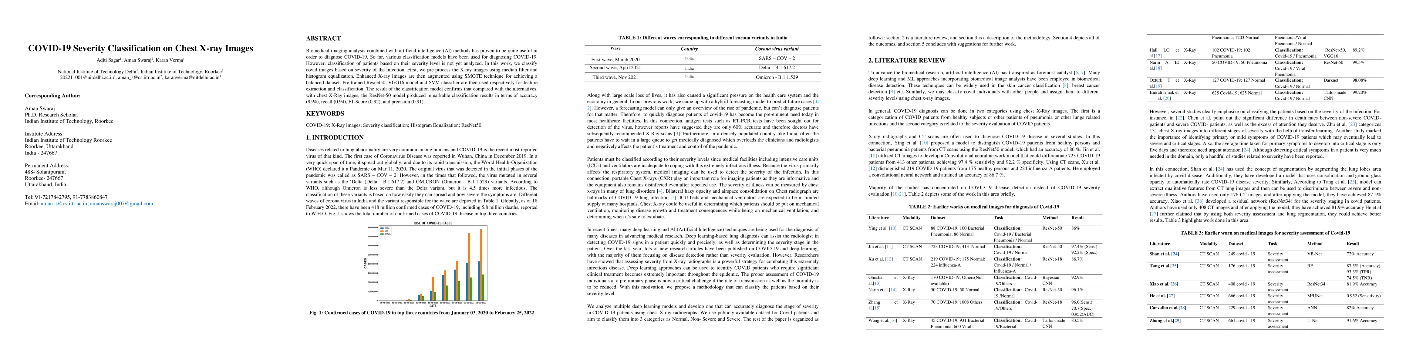
Biomedical imaging analysis combined with artificial intelligence (AI) methods has proven to be quite valuable in order to diagnose COVID-19. So far, various classification models have been used for diagnosing COVID-19. However, classification of patients based on their severity level is not yet analyzed. In this work, we classify covid images based on the severity of the infection. First, we pre-process the X-ray images using a median filter and histogram equalization. Enhanced X-ray images are then augmented using SMOTE technique for achieving a balanced dataset. Pre-trained Resnet50, VGG16 model and SVM classifier are then used for feature extraction and classification. The result of the classification model confirms that compared with the alternatives, with chest X-Ray images, the ResNet-50 model produced remarkable classification results in terms of accuracy (95%), recall (0.94), and F1-Score (0.92), and precision (0.91).
AI Key Findings
Get AI-generated insights about this paper's methodology, results, significance, and more — seven facets brought into focus.
Impact
Paper Details
Authors
PDF Preview
Key Terms
Citation Network
Current paper (gray), citations (green), references (blue)
Display is limited for performance on very large graphs.
Discussion 0