CovSegNet proposes a novel architecture for automatic lung lesion segmentation in COVID-19 chest CT scans, outperforming state-of-the-art approaches with minimal computational complexity.
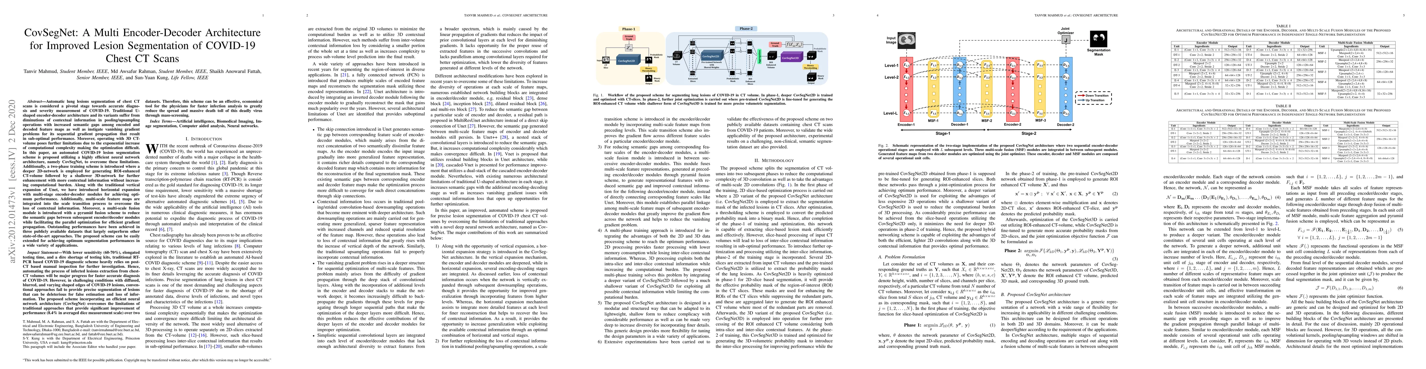
Automatic lung lesions segmentation of chest CT scans is considered a pivotal stage towards accurate diagnosis and severity measurement of COVID-19. Traditional U-shaped encoder-decoder architecture and its variants suffer from diminutions of contextual information in pooling/upsampling operations with increased semantic gaps among encoded and decoded feature maps as well as instigate vanishing gradient problems for its sequential gradient propagation that result in sub-optimal performance. Moreover, operating with 3D CT-volume poses further limitations due to the exponential increase of computational complexity making the optimization difficult. In this paper, an automated COVID-19 lesion segmentation scheme is proposed utilizing a highly efficient neural network architecture, namely CovSegNet, to overcome these limitations. Additionally, a two-phase training scheme is introduced where a deeper 2D-network is employed for generating ROI-enhanced CT-volume followed by a shallower 3D-network for further enhancement with more contextual information without increasing computational burden. Along with the traditional vertical expansion of Unet, we have introduced horizontal expansion with multi-stage encoder-decoder modules for achieving optimum performance. Additionally, multi-scale feature maps are integrated into the scale transition process to overcome the loss of contextual information. Moreover, a multi-scale fusion module is introduced with a pyramid fusion scheme to reduce the semantic gaps between subsequent encoder/decoder modules while facilitating the parallel optimization for efficient gradient propagation. Outstanding performances have been achieved in three publicly available datasets that largely outperform other state-of-the-art approaches. The proposed scheme can be easily extended for achieving optimum segmentation performances in a wide variety of applications.
Get AI-generated insights about this paper's methodology, results, and significance.
Current paper (gray), citations (green), references (blue)
Paschalis Bizopoulos, Nicholas Vretos, Petros Daras
Title | Authors | Year | Actions |
---|---|---|---|
Optimized attention U-Net for enhanced lung and area of infection segmentation in chest X-Rays and CT scans
|
Benzorgat Mustapha, Yatong Zhou, Benzorgat Nawel, Shan Chunyan, Zhitao Xiao
|
2025
|
View Paper |
TransGUNet: Transformer Meets Graph-based Skip Connection for Medical Image Segmentation
|
Ju-Hyeon Nam, Nur Suriza Syazwany, Sang-Chul Lee
|
2025
|
View Paper |
FIMKD: Feature-Implicit Mapping Knowledge Distillation for RGB-D Indoor Scene Semantic Segmentation
|
Wujie Zhou, Yuxiang Xiao, Yuanyuan Liu, Qiuping Jiang
|
2024
|
View Paper |
Multifunctional aggregation network of cell nuclei segmentation aiming histopathological diagnosis assistance: A new MA-Net construction
|
Qiumei Pu, Jinglong Tian, Donghao Wei, Qingming Shu, Minghui Sun, Lina Zhao
|
2024
|
View Paper |
Diverter transformer-based multi-encoder-multi-decoder network model for medical retinal blood vessel image segmentation
|
Chengwei Wu, Min Guo, Miao Ma, Kaiguang Wang
|
2024
|
View Paper |
Multiple Resolutions Detail Enhancement Network for Real-Time Image Semantic Segmentation
|
Jing Gu, Xinkai Sun, Jie Feng, Shuyuan Yang, Fang Liu, Licheng Jiao
|
2024
|
View Paper |
Improving 3-D Medical Image Segmentation at Boundary Regions Using Local Self-Attention and Global Volume Mixing
|
Daniya Najiha Abdul Kareem, M. Fiaz, Noa Novershtern, Jacob H. Hanna, Hisham Cholakkal
|
2024
|
View Paper |
Deep Learning-Based Face Mask Recognition System with YOLOv8
|
Christine Dewi, Danny Manongga, Hendry, Evangs Mailoa
|
2024
|
View Paper |
Online Aware Synapse Weighted Autoencoder for Recovering Random Missing Data in Wastewater Treatment Process
|
H. Han, Meiting Sun, Fang Li
|
2024
|
View Paper |
Deep Dual Attention Network for Precise Diagnosis of COVID-19 From Chest CT Images
|
Zhijie Lin, Zhaoshui He, Ruoyu Yao, Xu Wang, Taiheng Liu, Yamei Deng, Shengli Xie
|
2024
|
View Paper |
EdgeMedNet: Lightweight and Accurate U-Net for Implementing Efficient Medical Image Segmentation on Edge Devices
|
Qingliang Liu, Shuai Zhou, Jinmei Lai
|
2023
|
View Paper |
ERGPNet: lesion segmentation network for COVID-19 chest X-ray images based on embedded residual convolution and global perception
|
G. Yue, Chen Yang, Zhengyang Zhao, Ziheng An, Yongsheng Yang
|
2023
|
View Paper |
Critical Studies on Lesion Segmentation in Medical Images
|
Alok Kumar, N. Mahendran
|
2023
|
View Paper |
An Improved Grey Wolf Optimization–Based Convolutional Neural Network for the Segmentation of COVID-19 Lungs–Infected Parts
|
P. Sridhar, Jayaraj Ramasamy, Ravi Kumar, R. Ramanathan, R. Nayak, M. Tholkapiyan
|
2023
|
View Paper |
Automatic Medical Face Mask Recognition for COVID-19 Mitigation: Utilizing YOLO V5 Object Detection
|
Christine Dewi, Henoch Juli Christanto
|
2023
|
View Paper |
A systematic analysis and review of COVID-19 detection techniques using CT image
|
J. A. Beegom, Thomas Brindha
|
2023
|
View Paper |
YOLOv7 for Face Mask Identification Based on Deep Learning
|
Christine Dewi, Abbott Po Shun Chen, Henoch Juli Christanto
|
2023
|
View Paper |
Semi-Supervised Segmentation of COVID-19 Infection on CT-Scan Lung Using Dynamic Mutual Training
|
Ersa Resita, Tita Karlita, Riyanto Sigit, E. M. Yuniarno, I. K. Eddy, Purnama, M. Purnomo
|
2022
|
View Paper |
A Survey on Masked Facial Detection Methods and Datasets for Fighting Against COVID-19
|
Bingshu Wang, Jiangbin Zheng, C. L. P. Chen
|
2022
|
View Paper |
Rethinking Feature Guidance for Medical Image Segmentation
|
Wei Wang, Jixing He, Xin Wang
|
2025
|
View Paper |
A Spatially Aware Few-Shot Approach to Classification of Radar Sounder Data
|
Milkisa Yebasse, Lorenzo Bruzzone
|
2025
|
View Paper |
Machine learning-based anatomical segmentation: A systematic review of methodologies and applications
|
Evan Russell
|
View Paper |
Comments (0)