Cross Fusion and Correlation Beamformer for Row-Column Array Based 3D Ultrasound Imaging
Publication
Metrics
AI Quick Summary
The study introduces CFAC, a cross fusion and correlation method that reduces sidelobes and noise in row-column array 3D ultrasound, enabling higher contrast imaging. By using data from orthogonal apertures and multiple steering angles, CFAC markedly improves CNR in simulations, phantom, and in vivo rat kidney imaging while preserving ultrafast frame rates.
Paper Preview
Abstract
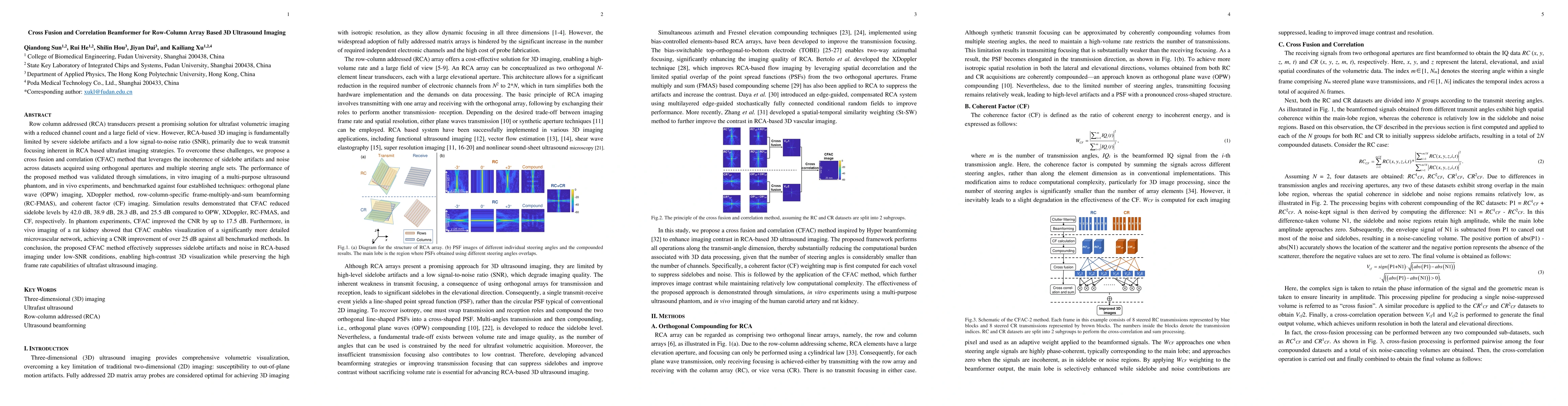
Row column addressed (RCA) transducers present a promising solution for ultrafast volumetric imaging with a reduced channel count and a large field of view. However, RCA-based 3D imaging is fundamentally limited by severe sidelobe artifacts and a low signal-to-noise ratio (SNR), primarily due to weak transmit focusing inherent in RCA based ultrafast imaging strategies. To overcome these challenges, we propose a cross fusion and correlation (CFAC) method that leverages the incoherence of sidelobe artifacts and noise across datasets acquired using orthogonal apertures and multiple steering angle sets. The performance of the proposed method was validated through simulations, in vitro imaging of a multi-purpose ultrasound phantom, and in vivo experiments, and benchmarked against four established techniques: orthogonal plane wave (OPW) imaging, XDoppler method, row-column-specific frame-multiply-and-sum beamforming (RC-FMAS), and coherent factor (CF) imaging. Simulation results demonstrated that CFAC reduced sidelobe levels by 42.0 dB, 38.9 dB, 28.3 dB, and 25.5 dB compared to OPW, XDoppler, RC-FMAS, and CF, respectively. In phantom experiments, CFAC improved the CNR by up to 17.5 dB. Furthermore, in vivo imaging of a rat kidney showed that CFAC enables visualization of a significantly more detailed microvascular network, achieving a CNR improvement of over 25 dB against all benchmarked methods. In conclusion, the proposed CFAC method effectively suppresses sidelobe artifacts and noise in RCA-based imaging under low-SNR conditions, enabling high-contrast 3D visualization while preserving the high frame rate capabilities of ultrafast ultrasound imaging.
AI Key Findings
Get AI-generated insights about this paper's methodology, results, significance, and more — seven facets brought into focus.
Discussion 0