Publication
Metrics
AI Quick Summary
This paper introduces a numerical algorithm for simulating cross-grating phase microscopy (CGM) data, allowing for the analysis of various experimental parameters' effects on noise, precision, and accuracy. The study aims to enhance CGM's application in biomicroscopy and nanophotonics by understanding the fundamental aspects of CGM.
Paper Preview
Abstract
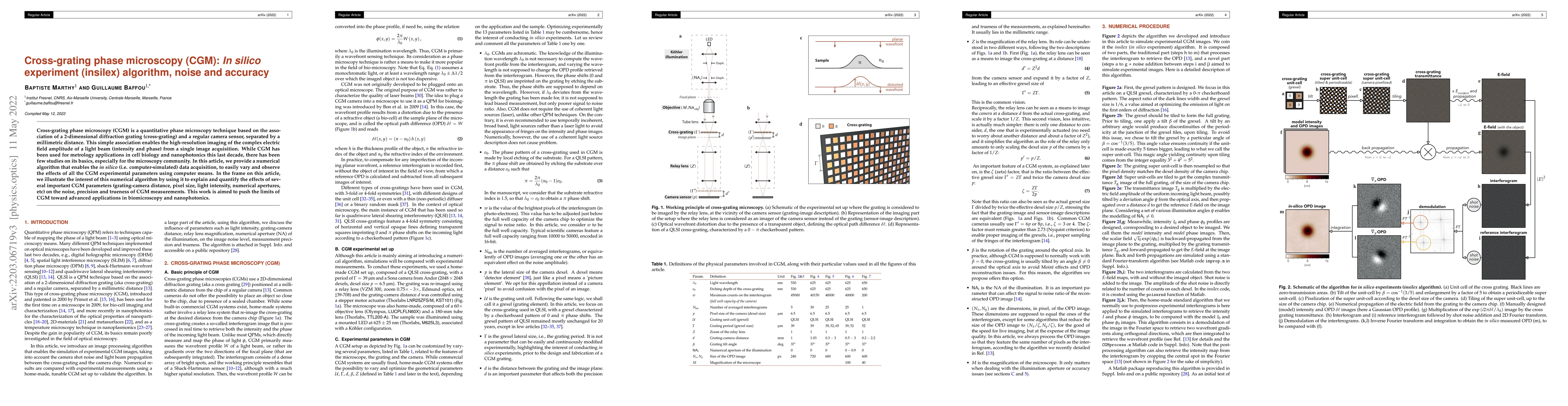
Cross-grating phase microscopy (CGM) is a quantitative phase microscopy technique based on the association of a 2-dimensional diffraction grating (cross-grating) and a regular camera sensor, separated by a millimetric distance. This simple association enables the high-resolution imaging of the complex electric field amplitude of a light beam (intensity and phase) from a single image acquisition. While CGM has been used for metrology applications in cell biology and nanophotonics this last decade, there has been few studies on its basics, especially for the microscopy community. In this article, we provide a numerical algorithm that enables the in silico (i.e. computer-simulated) data acquisition, to easily vary and observe the effects of all the CGM experimental parameters using computer means. In the frame on this article, we illustrate the interest of this numerical algorithm by using it to explain and quantify the effects of several important CGM parameters (grating-camera distance, pixel size, light intensity, numerical apertures, etc) on the noise, precision and trueness of CGM measurements. This work is aimed to push the limits of CGM toward advanced applications in biomicroscopy and nanophotonics.
AI Key Findings
Get AI-generated insights about this paper's methodology, results, significance, and more — seven facets brought into focus.
Impact
Paper Details
Authors
PDF Preview
Key Terms
Citation Network
Current paper (gray), citations (green), references (blue)
Display is limited for performance on very large graphs.
Discussion 0