Publication
Metrics
AI Quick Summary
This paper demonstrates the first experimental use of cryptotomography to reconstruct the 3D Fourier intensity distribution of nanoparticles from single-shot 2D diffraction patterns, addressing unmeasured photon fluence and data loss. This breakthrough extends the EMC framework and paves the way for single-shot diffraction imaging of biomolecules.
Paper Preview
Abstract
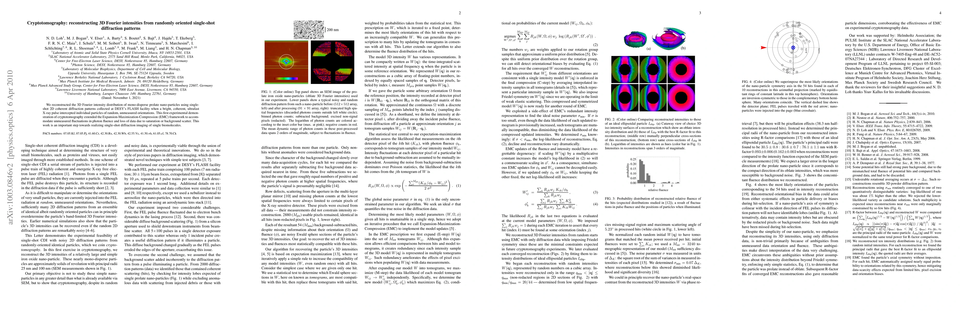
We reconstructed the 3D Fourier intensity distribution of mono-disperse prolate nano-particles using single-shot 2D coherent diffraction patterns collected at DESY's FLASH facility when a bright, coherent, ultrafast X-ray pulse intercepted individual particles of random, unmeasured orientations. This first experimental demonstration of cryptotomography extended the Expansion-Maximization-Compression (EMC) framework to accommodate unmeasured fluctuations in photon fluence and loss of data due to saturation or background scatter. This work is an important step towards realizing single-shot diffraction imaging of single biomolecules.
AI Key Findings
Get AI-generated insights about this paper's methodology, results, significance, and more — seven facets brought into focus.
Impact
Paper Details
PDF Preview
Key Terms
Citation Network
Current paper (gray), citations (green), references (blue)
Display is limited for performance on very large graphs.
Discussion 0