Summary
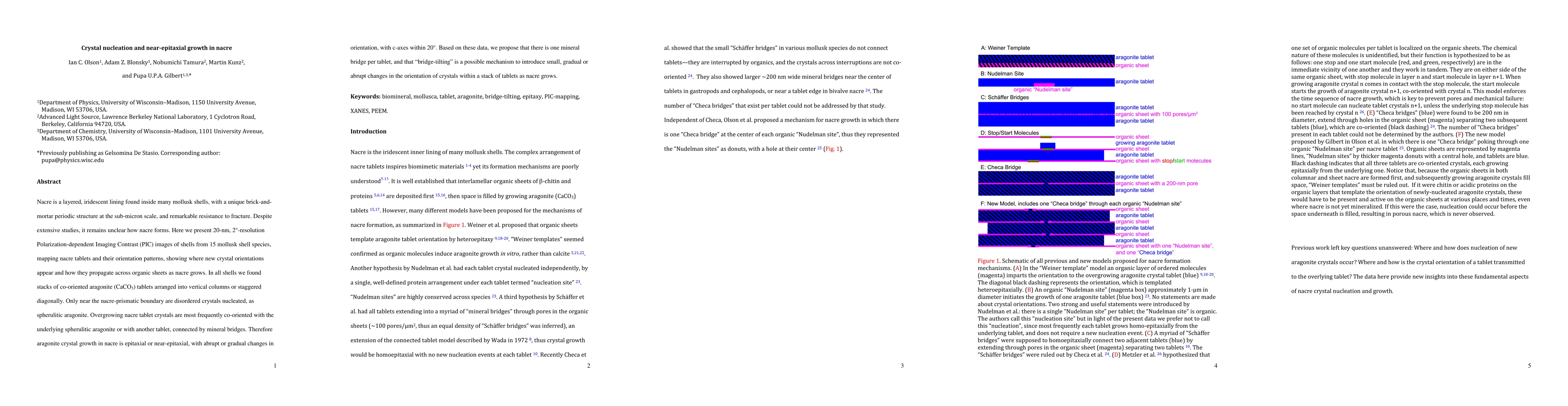
Nacre is a layered, iridescent lining found inside many mollusk shells, with a unique brick-and-mortar periodic structure at the sub-micron scale, and remarkable resistance to fracture. Despite extensive studies, it remains unclear how nacre forms. Here we present 20-nm, 2{\deg}-resolution Polarization-dependent Imaging Contrast (PIC) images of shells from 15 mollusk shell species, mapping nacre tablets and their orientation patterns, showing where new crystal orientations appear and how they propagate across organic sheets as nacre grows. In all shells we found stacks of co-oriented aragonite (CaCO3) tablets arranged into vertical columns or staggered diagonally. Only near the nacre-prismatic boundary are disordered crystals nucleated, as spherulitic aragonite. Overgrowing nacre tablet crystals are most frequently co-oriented with the underlying spherulitic aragonite or with another tablet, connected by mineral bridges. Therefore aragonite crystal growth in nacre is epitaxial or near-epitaxial, with abrupt or gradual changes in orientation, with c-axes within 20{\deg}. Based on these data, we propose that there is one mineral bridge per tablet, and that "bridge-tilting" is a possible mechanism to introduce small, gradual or abrupt changes in the orientation of crystals within a stack of tablets as nacre grows.
AI Key Findings
Get AI-generated insights about this paper's methodology, results, and significance.
Paper Details
PDF Preview
Key Terms
Citation Network
Current paper (gray), citations (green), references (blue)
Display is limited for performance on very large graphs.
Similar Papers
Found 4 papers| Title | Authors | Year | Actions |
|---|
Comments (0)