CT-based Anomaly Detection of Liver Tumors Using Generative Diffusion Prior
Publication
Metrics
AI Quick Summary
This research introduces a generative diffusion method for enhancing CT-based detection of liver tumors, improving upon traditional anomaly detection techniques. The method uses inpainting to refine liver references and anomaly scoring, showing a 7.9% increase in AUC compared to current methods, suggesting better radiological assessment.
Paper Preview
Abstract
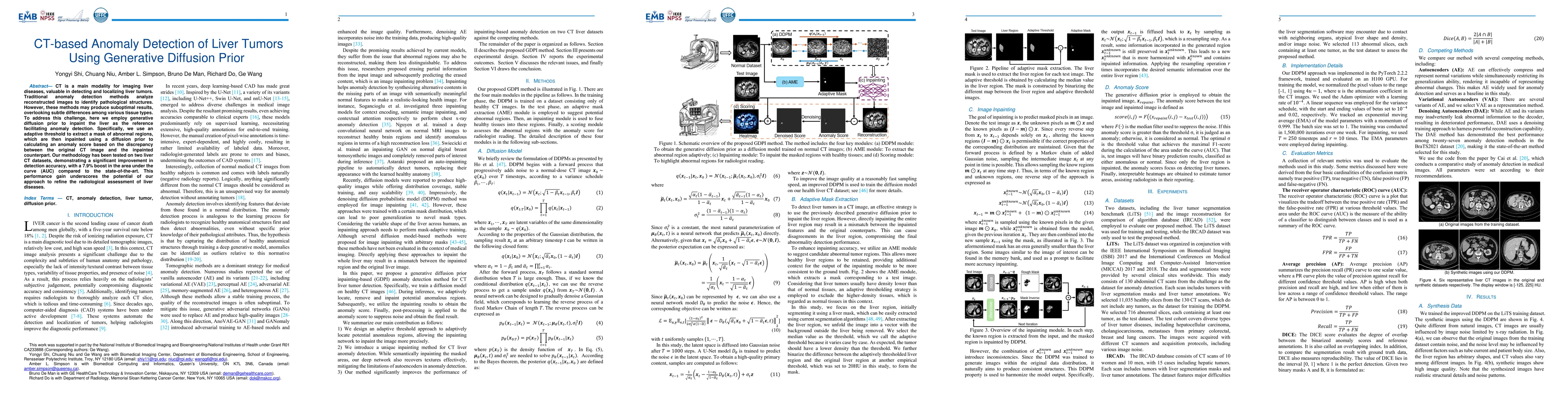
CT is a main modality for imaging liver diseases, valuable in detecting and localizing liver tumors. Traditional anomaly detection methods analyze reconstructed images to identify pathological structures. However, these methods may produce suboptimal results, overlooking subtle differences among various tissue types. To address this challenge, here we employ generative diffusion prior to inpaint the liver as the reference facilitating anomaly detection. Specifically, we use an adaptive threshold to extract a mask of abnormal regions, which are then inpainted using a diffusion prior to calculating an anomaly score based on the discrepancy between the original CT image and the inpainted counterpart. Our methodology has been tested on two liver CT datasets, demonstrating a significant improvement in detection accuracy, with a 7.9% boost in the area under the curve (AUC) compared to the state-of-the-art. This performance gain underscores the potential of our approach to refine the radiological assessment of liver diseases.
AI Key Findings
Get AI-generated insights about this paper's methodology, results, significance, and more — seven facets brought into focus.
Impact
Paper Details
Authors
PDF Preview
Citation Network
Current paper (gray), citations (green), references (blue)
Display is limited for performance on very large graphs.
Discussion 0