Authors
Summary
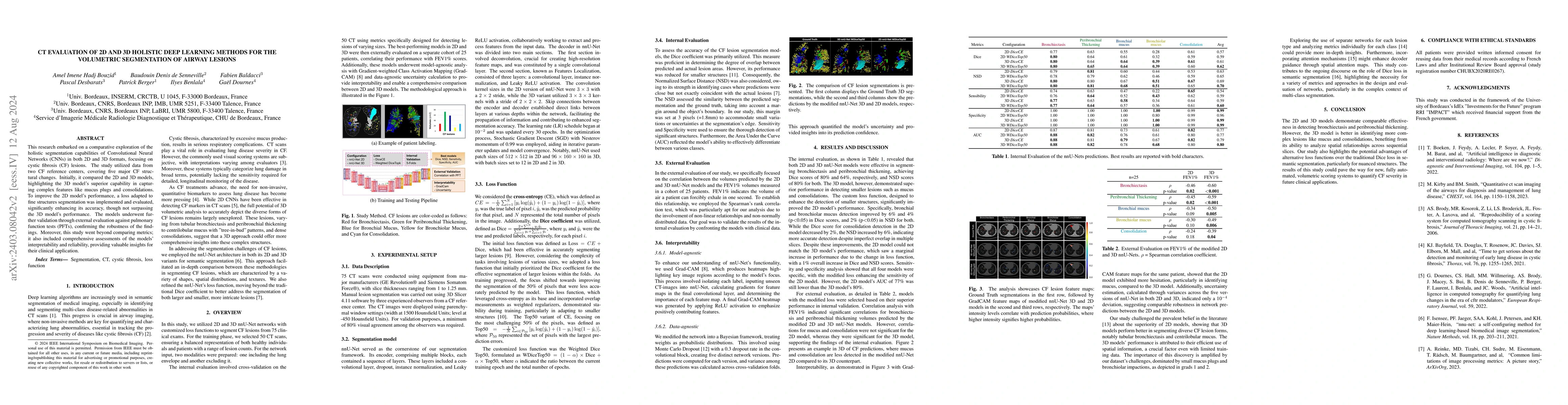
This research embarked on a comparative exploration of the holistic segmentation capabilities of Convolutional Neural Networks (CNNs) in both 2D and 3D formats, focusing on cystic fibrosis (CF) lesions. The study utilized data from two CF reference centers, covering five major CF structural changes. Initially, it compared the 2D and 3D models, highlighting the 3D model's superior capability in capturing complex features like mucus plugs and consolidations. To improve the 2D model's performance, a loss adapted to fine structures segmentation was implemented and evaluated, significantly enhancing its accuracy, though not surpassing the 3D model's performance. The models underwent further validation through external evaluation against pulmonary function tests (PFTs), confirming the robustness of the findings. Moreover, this study went beyond comparing metrics; it also included comprehensive assessments of the models' interpretability and reliability, providing valuable insights for their clinical application.
AI Key Findings
Get AI-generated insights about this paper's methodology, results, and significance.
Paper Details
PDF Preview
Key Terms
Citation Network
Current paper (gray), citations (green), references (blue)
Display is limited for performance on very large graphs.
Similar Papers
Found 4 papersBridging 2D and 3D Segmentation Networks for Computation Efficient Volumetric Medical Image Segmentation: An Empirical Study of 2.5D Solutions
Yichi Zhang, Jicong Zhang, Le Ding et al.
| Title | Authors | Year | Actions |
|---|
Comments (0)