Curved edges in the vertex model increase tissue fluidity
Publication
Metrics
AI Quick Summary
This paper explores the impact of curved edges in the Vertex Model on tissue fluidity, demonstrating that they lower the solid-to-fluid transition threshold from $p0 = 3.81$ to $p0 = 3.73$, thus enabling tissue to become more fluid at lower target perimeters.
Paper Preview
Abstract
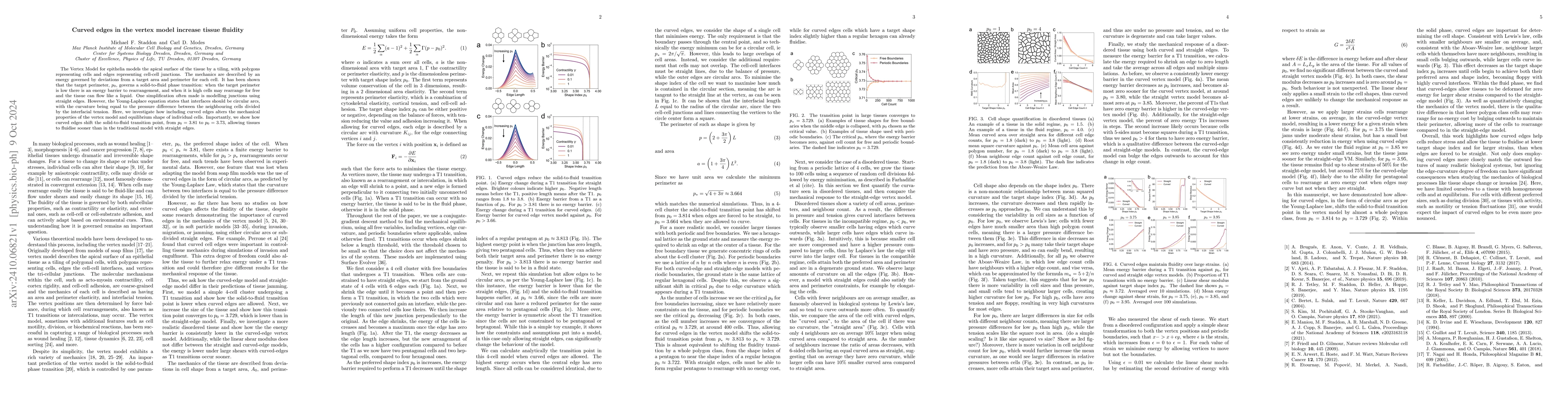
The Vertex Model for epithelia models the apical surface of the tissue by a tiling, with polygons representing cells and edges representing cell-cell junctions. The mechanics are described by an energy governed by deviations from a target area and perimeter for each cell. It has been shown that the target perimeter, p0, governs a solid-to-fluid phase transition: when the target perimeter is low there is an energy barrier to rearrangement, and when it is high cells may rearrange for free and the tissue can flow like a liquid. One simplification often made is modelling junctions using straight edges. However, the Young-Laplace equation states that interfaces should be circular arcs, with the curvature being equal to the pressure difference between the neighbouring cells divided by the interfacial tension. Here, we investigate how including curved edges alters the mechanical properties of the vertex model and equilibrium shape of individual cells. Importantly, we show how curved edges shift the solid-to-fluid transition point, from $p0 = 3.81$ to $p0 = 3.73$, allowing tissues to fluidise sooner than in the traditional model with straight edges.
AI Key Findings
Get AI-generated insights about this paper's methodology, results, significance, and more — seven facets brought into focus.
Impact
Authors
PDF Preview
Related Papers
No references found for this paper.
Discussion 0