Publication
Metrics
AI Quick Summary
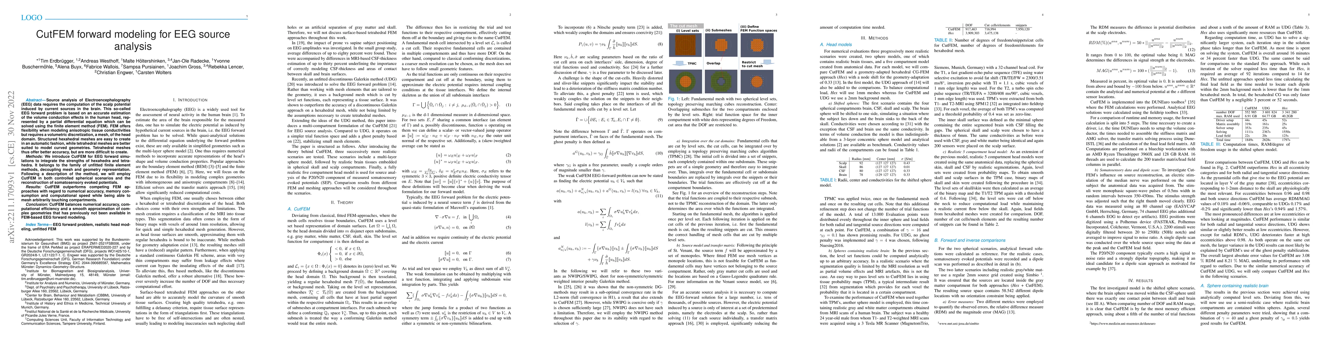
CutFEM forward modeling for EEG source analysis introduces an unfitted finite element method that combines the advantages of hexahedral and tetrahedral meshes for solving EEG forward problems, achieving higher accuracy, lower memory use, and faster computation compared to traditional FEM approaches. This method effectively handles complex geometries and outperforms existing methods in EEG simulations.
Paper Preview
Abstract
Source analysis of Electroencephalography (EEG) data requires the computation of the scalp potential induced by current sources in the brain. This so-called EEG forward problem is based on an accurate estimation of the volume conduction effects in the human head, represented by a partial differential equation which can be solved using the finite element method (FEM). FEM offers flexibility when modeling anisotropic tissue conductivities but requires a volumetric discretization, a mesh, of the head domain. Structured hexahedral meshes are easy to create in an automatic fashion, while tetrahedral meshes are better suited to model curved geometries. Tetrahedral meshes thus offer better accuracy, but are more difficult to create. Methods: We introduce CutFEM for EEG forward simulations to integrate the strengths of hexahedra and tetrahedra. It belongs to the family of unfitted finite element methods, decoupling mesh and geometry representation. Following a description of the method, we will employ CutFEM in both controlled spherical scenarios and the reconstruction of somatosensory evoked potentials. Results: CutFEM outperforms competing FEM approaches with regard to numerical accuracy, memory consumption and computational speed while being able to mesh arbitrarily touching compartments. Conclusion: CutFEM balances numerical accuracy, computational efficiency and a smooth approximation of complex geometries that has previously not been available in FEM-based EEG forward modeling.
AI Key Findings
Get AI-generated insights about this paper's methodology, results, significance, and more — seven facets brought into focus.
Impact
Paper Details
Authors
PDF Preview
Key Terms
Citation Network
Current paper (gray), citations (green), references (blue)
Display is limited for performance on very large graphs.
Discussion 0