Data Consistent CT Reconstruction from Insufficient Data with Learned Prior Images
Publication
Metrics
AI Quick Summary
This paper proposes a data consistent reconstruction (DCR) method to improve CT image reconstruction from insufficient data, combining deep learning and compressed sensing. The method generates a prior image, inpaints unmeasured projection data, and applies iterative reconstruction with regularization, achieving better accuracy and consistency compared to state-of-the-art U-Net methods.
Paper Preview
Abstract
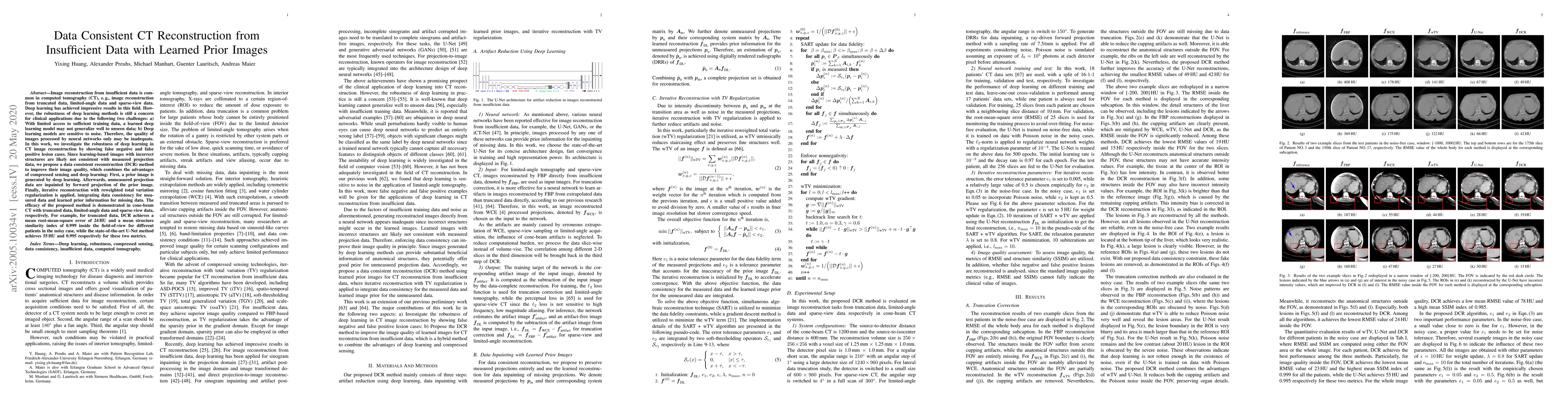
Image reconstruction from insufficient data is common in computed tomography (CT), e.g., image reconstruction from truncated data, limited-angle data and sparse-view data. Deep learning has achieved impressive results in this field. However, the robustness of deep learning methods is still a concern for clinical applications due to the following two challenges: a) With limited access to sufficient training data, a learned deep learning model may not generalize well to unseen data; b) Deep learning models are sensitive to noise. Therefore, the quality of images processed by neural networks only may be inadequate. In this work, we investigate the robustness of deep learning in CT image reconstruction by showing false negative and false positive lesion cases. Since learning-based images with incorrect structures are likely not consistent with measured projection data, we propose a data consistent reconstruction (DCR) method to improve their image quality, which combines the advantages of compressed sensing and deep learning: First, a prior image is generated by deep learning. Afterwards, unmeasured projection data are inpainted by forward projection of the prior image. Finally, iterative reconstruction with reweighted total variation regularization is applied, integrating data consistency for measured data and learned prior information for missing data. The efficacy of the proposed method is demonstrated in cone-beam CT with truncated data, limited-angle data and sparse-view data, respectively. For example, for truncated data, DCR achieves a mean root-mean-square error of 24 HU and a mean structure similarity index of 0.999 inside the field-of-view for different patients in the noisy case, while the state-of-the-art U-Net method achieves 55 HU and 0.995 respectively for these two metrics.
AI Key Findings
Get AI-generated insights about this paper's methodology, results, significance, and more — seven facets brought into focus.
Impact
Paper Details
Authors
PDF Preview
Key Terms
Citation Network
Current paper (gray), citations (green), references (blue)
Display is limited for performance on very large graphs.
Discussion 0