Decoding the mechanisms of phase transitions from in situ microscopy observations
Publication
Metrics
AI Quick Summary
Researchers used machine learning to analyze data from scanning electron microscopy images of BaTiO3 to visualize temperature-induced phase transitions. They found that the method can efficiently explore dynamic data and be applied to various imaging techniques and higher-dimensional spaces.
Paper Preview
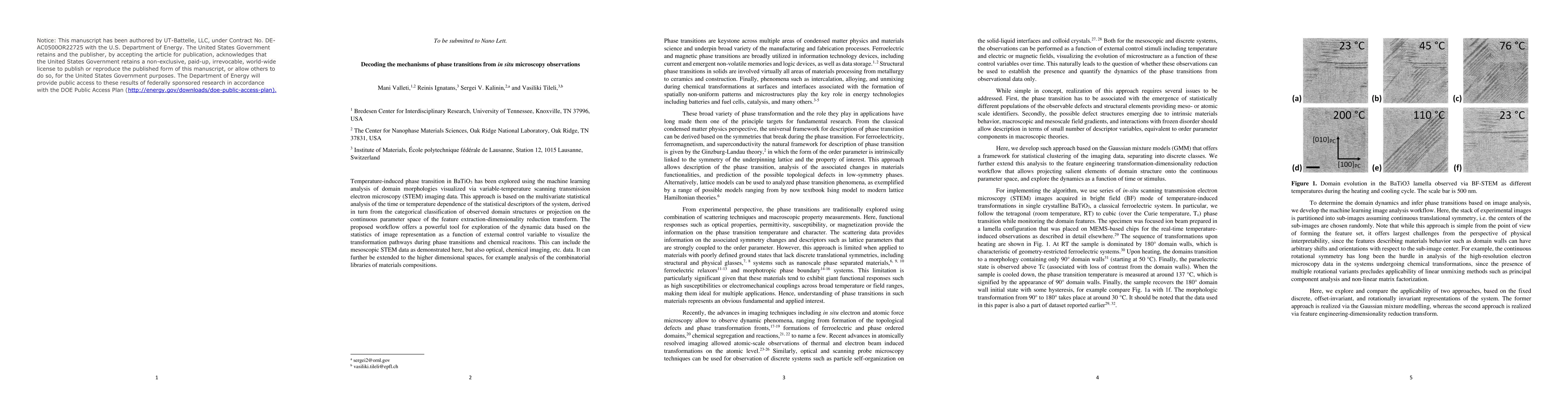
Abstract
Temperature-induced phase transition in BaTiO3 has been explored using the machine learning analysis of domain morphologies visualized via variable-temperature scanning transmission electron microscopy (STEM) imaging data. This approach is based on the multivariate statistical analysis of the time or temperature dependence of the statistical descriptors of the system, derived in turn from the categorical classification of observed domain structures or projection on the continuous parameter space of the feature extraction-dimensionality reduction transform. The proposed workflow offers a powerful tool for the exploration of the dynamic data based on the statistics of image representation as a function of the external control variable to visualize the transformation pathways during phase transitions and chemical reactions. This can include the mesoscopic STEM data as demonstrated here, but also optical, chemical imaging, etc. data. It can further be extended to the higher dimensional spaces, for example, analysis of the combinatorial libraries of materials compositions.
AI Key Findings
Get AI-generated insights about this paper's methodology, results, significance, and more — seven facets brought into focus.
Impact
Paper Details
Authors
PDF Preview
Key Terms
Citation Network
Current paper (gray), citations (green), references (blue)
Display is limited for performance on very large graphs.
Discussion 0