Researchers developed a new imaging technique that improves resolution of brain images, increasing clarity of cerebral vasculature by 1.9 times, with applications for further brain research.
We proposed a deconvolution optical-resolution photoacoustic microscope for high -resolution imaging of brain. The focal spot of the photoacoustic microscopy is measured to obtain the lateral PSF (point spread function) of the system. Making the measured PSF as the initial system PSF to perform Lucy- Richardson (LR) deconvolution. The image resolution of cerebral vasculature obtained by this method is higher. The full width at half maximum (FWHM) of the width of blood vessel before and after deconvolution are 7 micrometers and 3.6 micrometers, respectively, and the image definition is increased by about 1.9 times. Experiments show that this method can further improve the clarity of photoacoustic images of cerebral vasculature, which lays the foundation for further research on brain imaging.
Generated Sep 03, 2025
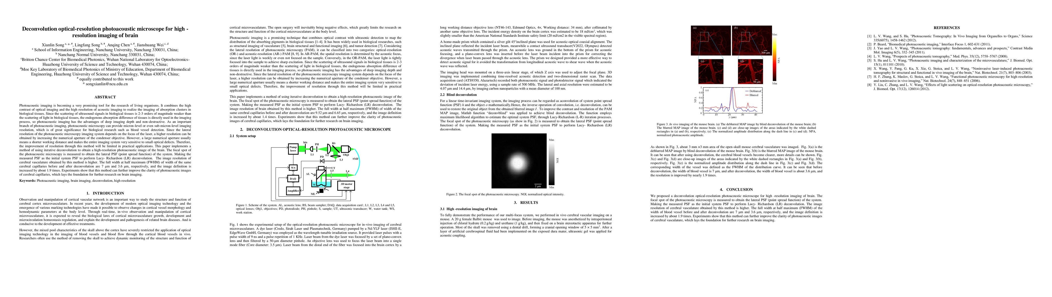
The research proposes a deconvolution optical-resolution photoacoustic microscope (OR-PAM) for high-resolution imaging of the brain. It measures the focal spot of the photoacoustic microscopy to obtain the lateral point spread function (PSF) of the system, which is then used as the initial system PSF for Lucy-Richardson (LR) deconvolution to enhance image resolution.
This research is significant as it improves the clarity of photoacoustic images of cerebral vasculature, which is crucial for further brain imaging studies, potentially aiding in understanding cortical microvasculature structure and function without the negative effects of open surgery.
The main technical contribution is the implementation of iterative deconvolution using Lucy-Richardson algorithm to obtain high-resolution photoacoustic images of the brain, utilizing the measured focal spot of the photoacoustic microscopy to determine the system's PSF.
This work introduces an innovative approach to enhance the resolution of photoacoustic microscopy images by employing deconvolution techniques, specifically Lucy-Richardson algorithm, which has not been extensively explored in brain imaging applications.
Current paper (gray), citations (green), references (blue)
Display is limited for performance on very large graphs.
Title | Authors | Year | Actions |
---|
Comments (0)