Publication
Metrics
AI Quick Summary
The paper proposes a Decoupled Pyramid Correlation Network (DPC-Net) for liver tumor segmentation in CT images, utilizing a Pyramid Feature Encoder and attention mechanisms to enhance both low- and high-level features. The method achieves superior segmentation results with a Dice Similarity Coefficient of 76.4% and an Average Symmetric Surface Distance of 0.838 mm, outperforming existing state-of-the-art techniques.
Paper Preview
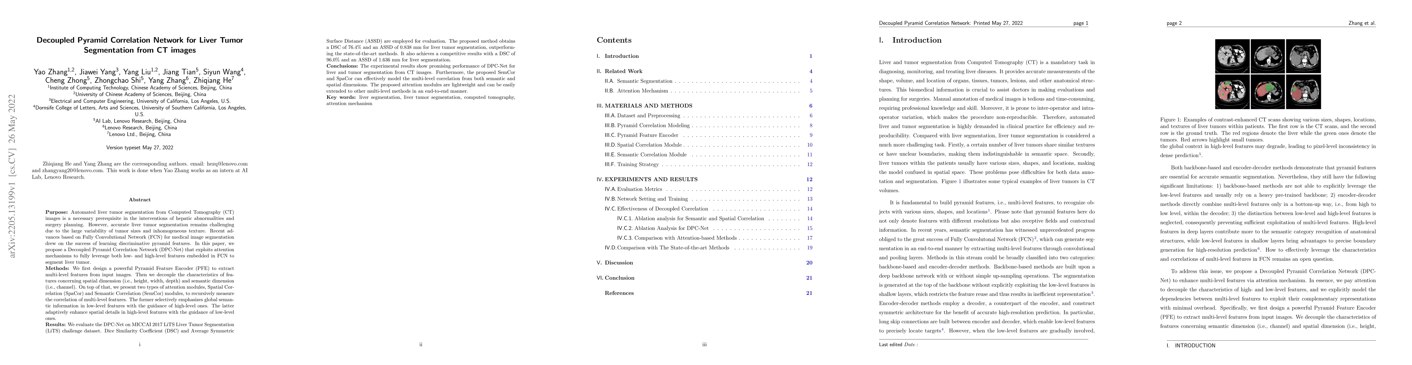
Abstract
Purpose: Automated liver tumor segmentation from Computed Tomography (CT) images is a necessary prerequisite in the interventions of hepatic abnormalities and surgery planning. However, accurate liver tumor segmentation remains challenging due to the large variability of tumor sizes and inhomogeneous texture. Recent advances based on Fully Convolutional Network (FCN) for medical image segmentation drew on the success of learning discriminative pyramid features. In this paper, we propose a Decoupled Pyramid Correlation Network (DPC-Net) that exploits attention mechanisms to fully leverage both low- and high-level features embedded in FCN to segment liver tumor. Methods: We first design a powerful Pyramid Feature Encoder (PFE) to extract multi-level features from input images. Then we decouple the characteristics of features concerning spatial dimension (i.e., height, width, depth) and semantic dimension (i.e., channel). On top of that, we present two types of attention modules, Spatial Correlation (SpaCor) and Semantic Correlation (SemCor) modules, to recursively measure the correlation of multi-level features. The former selectively emphasizes global semantic information in low-level features with the guidance of high-level ones. The latter adaptively enhance spatial details in high-level features with the guidance of low-level ones. Results: We evaluate the DPC-Net on MICCAI 2017 LiTS Liver Tumor Segmentation (LiTS) challenge dataset. Dice Similarity Coefficient (DSC) and Average Symmetric Surface Distance (ASSD) are employed for evaluation. The proposed method obtains a DSC of 76.4% and an ASSD of 0.838 mm for liver tumor segmentation, outperforming the state-of-the-art methods. It also achieves a competitive results with a DSC of 96.0% and an ASSD of 1.636 mm for liver segmentation.
AI Key Findings
Get AI-generated insights about this paper's methodology, results, significance, and more — seven facets brought into focus.
Impact
Paper Details
Authors
PDF Preview
Key Terms
Citation Network
Current paper (gray), citations (green), references (blue)
Display is limited for performance on very large graphs.
Discussion 0