Deep CNN ensembles and suggestive annotations for infant brain MRI segmentation
Publication
Metrics
AI Quick Summary
This study explores the use of an ensemble of semi-dense fully convolutional neural networks (CNNs) for precise 3D segmentation of infant brain tissues in MRI, addressing the challenges of poor image quality and isointense contrast. The proposed SemiDenseNet architecture connects all convolutional layers directly, significantly reducing parameters and improving gradient propagation. The ensemble's agreement correlates with segmentation errors, providing guidance for local user corrections. The method was evaluated on the MICCAI iSEG-2017 Challenge dataset, achieving competitive results among 21 teams.
Paper Preview
Abstract
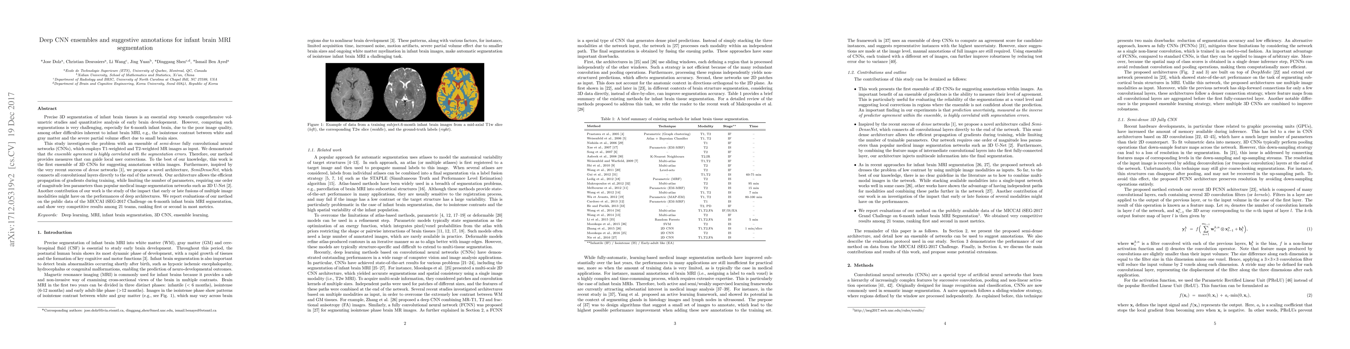
Precise 3D segmentation of infant brain tissues is an essential step towards comprehensive volumetric studies and quantitative analysis of early brain developement. However, computing such segmentations is very challenging, especially for 6-month infant brain, due to the poor image quality, among other difficulties inherent to infant brain MRI, e.g., the isointense contrast between white and gray matter and the severe partial volume effect due to small brain sizes. This study investigates the problem with an ensemble of semi-dense fully convolutional neural networks (CNNs), which employs T1-weighted and T2-weighted MR images as input. We demonstrate that the ensemble agreement is highly correlated with the segmentation errors. Therefore, our method provides measures that can guide local user corrections. To the best of our knowledge, this work is the first ensemble of 3D CNNs for suggesting annotations within images. Furthermore, inspired by the very recent success of dense networks, we propose a novel architecture, SemiDenseNet, which connects all convolutional layers directly to the end of the network. Our architecture allows the efficient propagation of gradients during training, while limiting the number of parameters, requiring one order of magnitude less parameters than popular medical image segmentation networks such as 3D U-Net. Another contribution of our work is the study of the impact that early or late fusions of multiple image modalities might have on the performances of deep architectures. We report evaluations of our method on the public data of the MICCAI iSEG-2017 Challenge on 6-month infant brain MRI segmentation, and show very competitive results among 21 teams, ranking first or second in most metrics.
AI Key Findings
Get AI-generated insights about this paper's methodology, results, significance, and more — seven facets brought into focus.
Impact
Paper Details
PDF Preview
Key Terms
Citation Network
Current paper (gray), citations (green), references (blue)
Display is limited for performance on very large graphs.
Discussion 0