ArxivLens

Quick Summary:

This paper introduces Deep-FExt, a machine learning-based feature extraction method using inception models for vessel segmentation and centerline prediction in biomedical images. The proposed method outperforms existing feature extraction techniques, achieving an average Dice coefficient of 0.85 on the DRIVE and STARE datasets, and extends the approach for potential 3-D dataset applications.

Journal Reference: Wang Q., Shi Y., Suk HI., Suzuki K. (eds) Machine Learning in Medical Imaging. MLMI 2017. Lecture Notes in Computer Science, vol 10541. Springer, Cham
DOI: 10.1007/978-3-319-67389-9_40
arXiv Id: 1704.03743
Comments: 9 pages
Report No: pp 344-352
Date Published: 2018-04-09
Date Updated: 2018-04-09

Categories:

Key Terms (TF-IDF):

feature extraction (0.288)
extraction (0.239)
data set (0.231)
feature (0.209)
pixel (0.197)
drive (0.187)
performs (0.178)
classification (0.176)
convolutional (0.165)
schemes (0.164)
purpose (0.164)
layers (0.158)
maximum (0.143)
end (0.137)
average (0.136)
scheme (0.133)
achieve (0.133)
image (0.132)
set (0.132)
datasets (0.128)
License: http://arxiv.org/licenses/nonexclusive-distrib/1.0/

Summary:

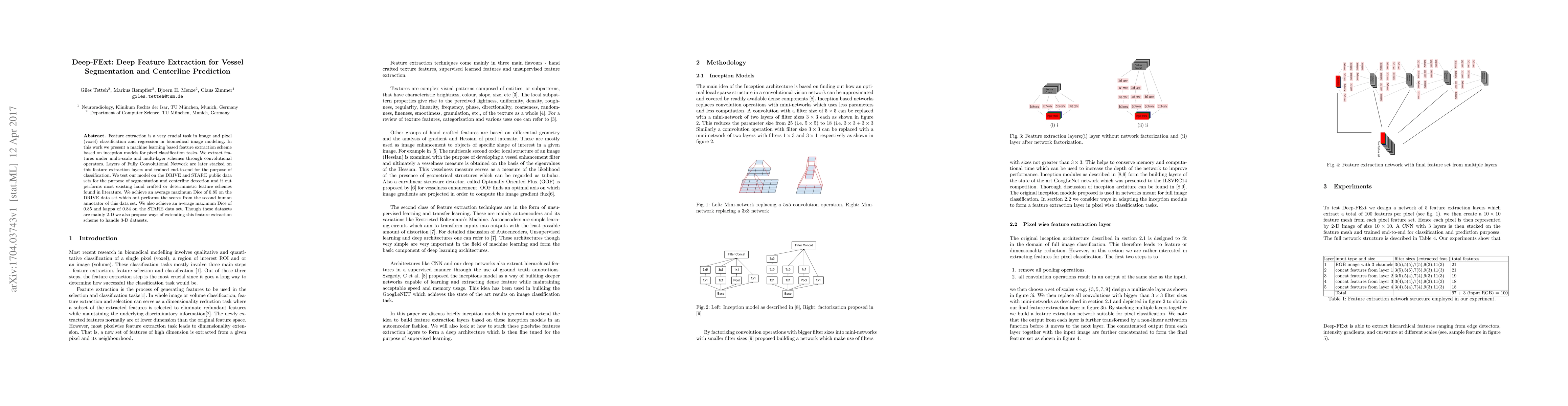
Feature extraction is a very crucial task in image and pixel (voxel) classification and regression in biomedical image modeling. In this work we present a machine learning based feature extraction scheme based on inception models for pixel classification tasks. We extract features under multi-scale and multi-layer schemes through convolutional operators. Layers of Fully Convolutional Network are later stacked on this feature extraction layers and trained end-to-end for the purpose of classification. We test our model on the DRIVE and STARE public data sets for the purpose of segmentation and centerline detection and it out performs most existing hand crafted or deterministic feature schemes found in literature. We achieve an average maximum Dice of 0.85 on the DRIVE data set which out performs the scores from the second human annotator of this data set. We also achieve an average maximum Dice of 0.85 and kappa of 0.84 on the STARE data set. Though these datasets are mainly 2-D we also propose ways of extending this feature extraction scheme to handle 3-D datasets.

Citation & Reference Network Visualization

This graph shows how this paper is connected to other research. The current paper is shown in dark gray, papers that cite it are in green, and papers it references are in blue. You can:

Current Paper
Papers citing this work (Citations)
Papers cited by this work (References)

• Hover over any node to see the paper title

• Click a node to view that paper's details

• Drag nodes to rearrange the graph

• Use the zoom controls (bottom right) to zoom in/out

Citations and References

Title Authors Year Actions
A comprehensive survey on segmentation techniques for retinal vessel segmentation
Jair Cervantes, Jared Cervantes, Farid García, Arturo Yee Rendón, Josué Espejel Cabrera, Laura D. Jalili
2023
View Paper
Ordered multi-path propagation for vessel centerline extraction
Tao Han, Danni Ai, Ruirui An, Jingfan Fan, Hong Song, Yining Wang, Jian Yang
2021
View Paper
Learning-Based Algorithms for Vessel Tracking: A Review
Dengqiang Jia, X. Zhuang
2020
View Paper
Convolutional Neural Network Ensemble Segmentation With Ratio-Based Sampling for the Arteries and Veins in Abdominal CT Scans
Alena-Kathrin Golla, Dominik F. Bauer, R. Schmidt, Tom Russ, D. Nörenberg, Khanlian Chung, Christian Tönnes, L. Schad, F. Zöllner
2020
View Paper
Encoding anatomical tree priors for tubular structure extraction for medical images analysis
Mengliu Zhao
2020
View Paper
Inception Capsule Network for Retinal Blood Vessel Segmentation and Centerline Extraction
Christian Kromm, K. Rohr
2019
View Paper
Unsupervised Ensemble Strategy for Retinal Vessel Segmentation
Bo Liu, Lin Gu, Feng Lu
2019
View Paper
Scale-space approximated convolutional neural networks for retinal vessel segmentation
Kyoung Jin Noh, S. Park, Soochahn Lee
2019
View Paper
DeepVesselNet: Vessel Segmentation, Centerline Prediction, and Bifurcation Detection in 3-D Angiographic Volumes
Giles Tetteh, Velizar Efremov, N. Forkert, Matthias Schneider, J. Kirschke, B. Weber, C. Zimmer, M. Piraud, Bjoern H Menze
2018
View Paper
Frangi-Net
Weilin Fu, Katharina Breininger, Roman Schaffert, N. Ravikumar, Tobias Würfl, Jim Fujimoto, Eric M. Moult, A. Maier
2017
View Paper
From Large to Small Organ Segmentation in CT Using Regional Context
M. Bieth, Esther Alberts, M. Schwaiger, Bjoern H Menze
2017
View Paper
A Survey of Deep Learning for Retinal Blood Vessel Segmentation Methods: Taxonomy, Trends, Challenges and Future Directions
O. Sule
2022
View Paper
BLU-GAN: Bi-directional ConvLSTM U-Net with Generative Adversarial Training for Retinal Vessel Segmentation
Li Lin, Jiewei Wu, Pujin Cheng, Kai Wang, Xiaoying Tang
2021
View Paper
Intracranial Vascular Structure Extraction: A Machine Learning Approach
Shi-feng Zhao, Yun Tian, Xuesong Wang, Lizhi Xie, Lingling Sun
2019
View Paper

Similar Papers

An automated framework for brain vessel centerline extraction from CTA images

Nanning Zheng, Ruisheng Su, Jingmin Xin, Jiayi Wu, Aad van der Lugt, Theo van Walsum, Wiro J. Niessen, Sijie Liu, Jianghang Su, Wim van Zwam, Pieter Jan van Doormaal

Published: 2024-01-17

Graph Convolution Based Cross-Network Multi-Scale Feature Fusion for Deep Vessel Segmentation

Yizhou Yu, Kongming Liang, Chengwei Pan, Gangming Zhao, Fandong Zhang, Xinyang Hu, Xianpeng Wu

Published: 2023-01-09

Comments (0)

This website uses cookies to ensure you get the best experience. Learn more