Deep ICE: A Deep learning approach for MRI Intracranial Cavity Extraction
Publication
Metrics
Paper Preview
Abstract
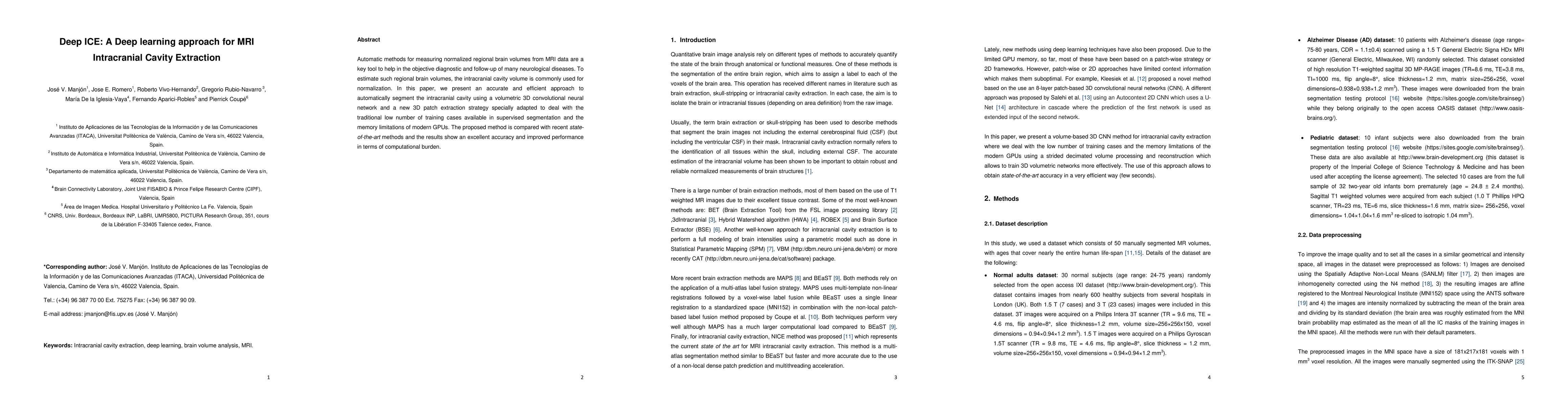
Automatic methods for measuring normalized regional brain volumes from MRI data are a key tool to help in the objective diagnostic and follow-up of many neurological diseases. To estimate such regional brain volumes, the intracranial cavity volume is commonly used for normalization. In this paper, we present an accurate and efficient approach to automatically segment the intracranial cavity using a volumetric 3D convolutional neural network and a new 3D patch extraction strategy specially adapted to deal with the traditional low number of training cases available in supervised segmentation and the memory limitations of modern GPUs. The proposed method is compared with recent state-of-the-art methods and the results show an excellent accuracy and improved performance in terms of computational burden.
AI Key Findings
Get AI-generated insights about this paper's methodology, results, significance, and more — seven facets brought into focus.
Impact
Paper Details
Authors
PDF Preview
Key Terms
Citation Network
Current paper (gray), citations (green), references (blue)
Display is limited for performance on very large graphs.
Discussion 0