Summary
Glaucoma is the leading cause of preventable, irreversible blindness world-wide. The disease can remain asymptomatic until severe, and an estimated 50%-90% of people with glaucoma remain undiagnosed. Glaucoma screening is recommended for early detection and treatment. A cost-effective tool to detect glaucoma could expand screening access to a much larger patient population, but such a tool is currently unavailable. We trained a deep learning algorithm using a retrospective dataset of 86,618 images, assessed for glaucomatous optic nerve head features and referable glaucomatous optic neuropathy (GON). The algorithm was validated using 3 datasets. For referable GON, the algorithm had an AUC of 0.945 (95% CI, 0.929-0.960) in dataset A (1205 images, 1 image/patient; 18.1% referable), images adjudicated by panels of Glaucoma Specialists (GSs); 0.855 (95% CI, 0.841-0.870) in dataset B (9642 images, 1 image/patient; 9.2% referable), images from Atlanta Veterans Affairs Eye Clinic diabetic teleretinal screening program; and 0.881 (95% CI, 0.838-0.918) in dataset C (346 images, 1 image/patient; 81.7% referable), images from Dr. Shroff's Charity Eye Hospital's glaucoma clinic. The algorithm showed significantly higher sensitivity than 7 of 10 graders not involved in determining the reference standard, including 2 of 3 GSs, and showed higher specificity than 3 graders, while remaining comparable to others. For both GSs and the algorithm, the most crucial features related to referable GON were: presence of vertical cup-to-disc ratio of 0.7 or more, neuroretinal rim notching, retinal nerve fiber layer defect, and bared circumlinear vessels. An algorithm trained on fundus images alone can detect referable GON with higher sensitivity than and comparable specificity to eye care providers. The algorithm maintained good performance on an independent dataset with diagnoses based on a full glaucoma workup.
AI Key Findings
Get AI-generated insights about this paper's methodology, results, and significance.
Paper Details
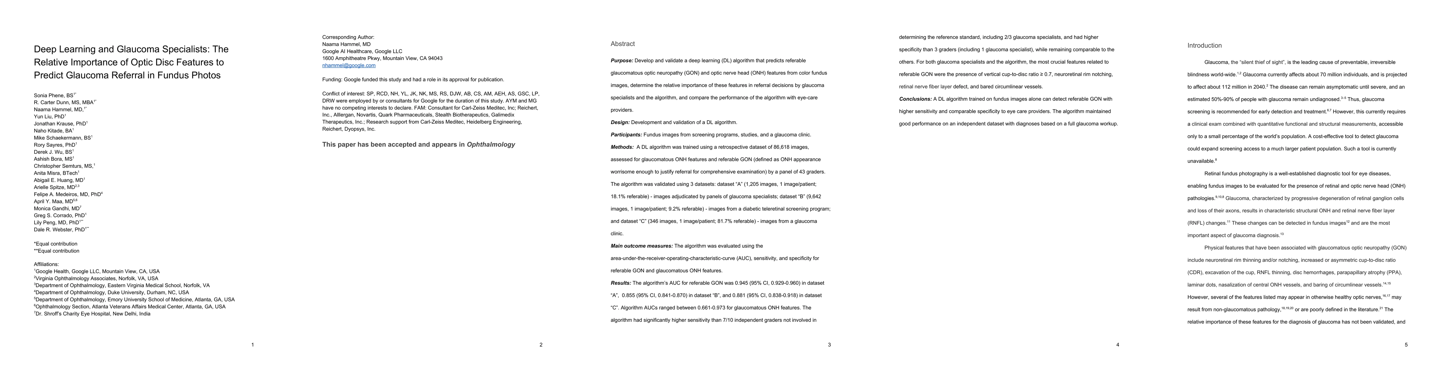
PDF Preview
Key Terms
Citation Network
Current paper (gray), citations (green), references (blue)
Display is limited for performance on very large graphs.
Similar Papers
Found 4 papers| Title | Authors | Year | Actions |
|---|
Comments (0)