The study trains a vision‑transformer based deep‑learning model on low‑resolution pre‑scan thumbnails extracted from whole‑slide images, using a tiled‑slide classification pipeline with soft‑voting aggregation. The model is trained on 1,200 Leica‑GT450DX WSIs from TUM and evaluated on class‑balanced external datasets (TCGA, Augsburg, Regensburg).
Deep Learning-Based Fixation Type Prediction for Quality Assurance in Digital Pathology
Publication
Metrics
Quick Answers
What methodology did the authors use?
The study trains a vision‑transformer based deep‑learning model on low‑resolution pre‑scan thumbnails extracted from whole‑slide images, using a tiled‑slide classification pipeline with soft‑voting aggregation. The model is trained on 1,200 Leica‑GT450DX WSIs from TUM and evaluated on class‑balanced external datasets (TCGA, Augsburg, Regensburg). More in Methodology →
What are the key results?
Achieves an AUROC of 0.88 on the TCGA dataset, surpassing prior thumbnail‑based methods by 4.8 % and matching the performance of full‑resolution approaches while being 400× faster. The best configuration (UNI backbone with soft‑voting) reaches 89 % accuracy on the TUM test set and processes each slide in 21 ms. Performance drops to 56 % accuracy on Philips slides and 50 % on 3DHISTECH slides,... More in Key Results →
Why is this work significant?
This work demonstrates that fixation‑type prediction can be performed rapidly and accurately using only pre‑scan thumbnails, enabling high‑throughput quality control in digital pathology without the need for costly full‑resolution scans. The speed‑accuracy trade‑off makes it practical for routine laboratory workflows and could be extended to other slide‑level annotations. More in Significance →
What are the main limitations?
The model’s performance degrades on slides from non‑Leica scanners, indicating a strong scanner‑dependent domain shift. The study does not address stain or color calibration variations across institutions. More in Limitations →
Abstract
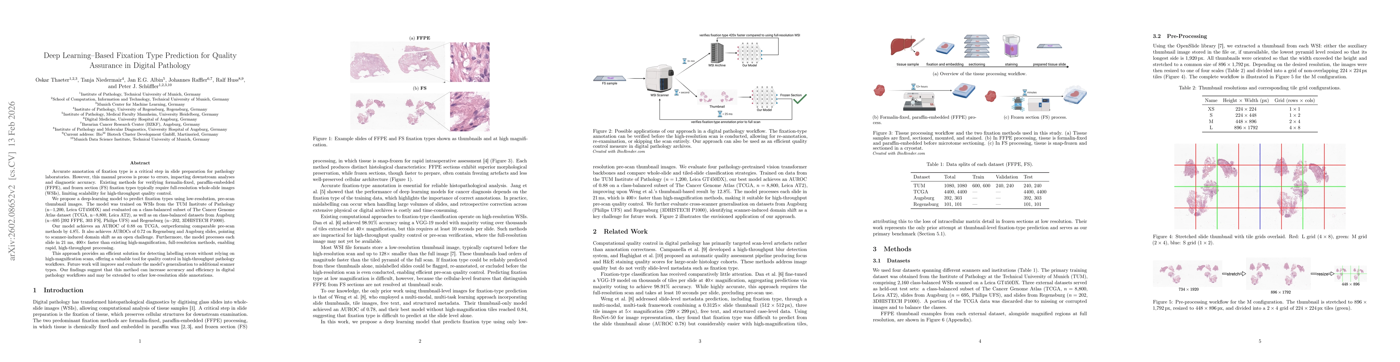
Accurate annotation of fixation type is a critical step in slide preparation for pathology laboratories. However, this manual process is prone to errors, impacting downstream analyses and diagnostic accuracy. Existing methods for verifying formalin-fixed, paraffin-embedded (FFPE), and frozen section (FS) fixation types typically require full-resolution whole-slide images (WSIs), limiting scalability for high-throughput quality control. We propose a deep-learning model to predict fixation types using low-resolution, pre-scan thumbnail images. The model was trained on WSIs from the TUM Institute of Pathology (n=1,200, Leica GT450DX) and evaluated on a class-balanced subset of The Cancer Genome Atlas dataset (TCGA, n=8,800, Leica AT2), as well as on class-balanced datasets from Augsburg (n=695 [392 FFPE, 303 FS], Philips UFS) and Regensburg (n=202, 3DHISTECH P1000). Our model achieves an AUROC of 0.88 on TCGA, outperforming comparable pre-scan methods by 4.8%. It also achieves AUROCs of 0.72 on Regensburg and Augsburg slides, underscoring challenges related to scanner-induced domain shifts. Furthermore, the model processes each slide in 21 ms, $400\times$ faster than existing high-magnification, full-resolution methods, enabling rapid, high-throughput processing. This approach provides an efficient solution for detecting labelling errors without relying on high-magnification scans, offering a valuable tool for quality control in high-throughput pathology workflows. Future work will improve and evaluate the model's generalisation to additional scanner types. Our findings suggest that this method can increase accuracy and efficiency in digital pathology workflows and may be extended to other low-resolution slide annotations.
Key Findings, in focus
Seven facets of this paper, analysed and brought into focus by AI.
This work demonstrates that fixation‑type prediction can be performed rapidly and accurately using only pre‑scan thumbnails, enabling high‑throughput quality control in digital pathology without the need for costly full‑resolution scans.
- Achieves an AUROC of 0.88 on the TCGA dataset, surpassing prior thumbnail‑based methods by 4.8 % and matching the performance of full‑resolution approaches while being 400× faster. The best configuration (UNI backbone with soft‑voting) reaches 89 % accuracy on the TUM test set and processes each slide in 21 ms. Performance drops to 56 % accuracy on Philips slides and 50 % on 3DHISTECH slides, highlighting scanner‑dependent domain shift.
This work demonstrates that fixation‑type prediction can be performed rapidly and accurately using only pre‑scan thumbnails, enabling high‑throughput quality control in digital pathology without the need for costly full‑resolution scans. The speed‑accuracy trade‑off makes it practical for routine laboratory workflows and could be extended to other slide‑level annotations.
The paper introduces the first efficient, thumbnail‑only fixation‑type classifier that leverages vision‑transformer backbones with tiled soft‑voting, achieving state‑of‑the‑art AUROC while reducing inference time to 21 ms per slide.
Unlike prior work that relies on high‑magnification patches, this study shows that low‑resolution thumbnails suffice for accurate fixation prediction, combining transformer‑based feature extraction with a lightweight aggregation strategy to achieve unprecedented speed and accuracy in pre‑scan quality control.
- The model’s performance degrades on slides from non‑Leica scanners, indicating a strong scanner‑dependent domain shift. The study does not address stain or color calibration variations across institutions.
- Apply domain adaptation techniques such as stain normalization, scanner‑aware training, or multi‑site fine‑tuning to improve cross‑scanner generalization. Incorporate additional pre‑scan modalities (e.g., barcode metadata, macro images) and develop explainability methods to support clinical adoption.
Discussion 0