Deep learning based projection domain metal segmentation for metal artifact reduction in cone beam computed tomography
Publication
Metrics
AI Quick Summary
This paper proposes a deep learning method for metal segmentation in cone beam computed tomography (CBCT) to reduce metal artifacts, using synthetic training data generated from X-ray simulations. The study shows that a small number of photons in simulations and training with both full and cropped projections improve model robustness, leading to significant improvements in image quality for various artifact conditions.
Paper Preview
Abstract
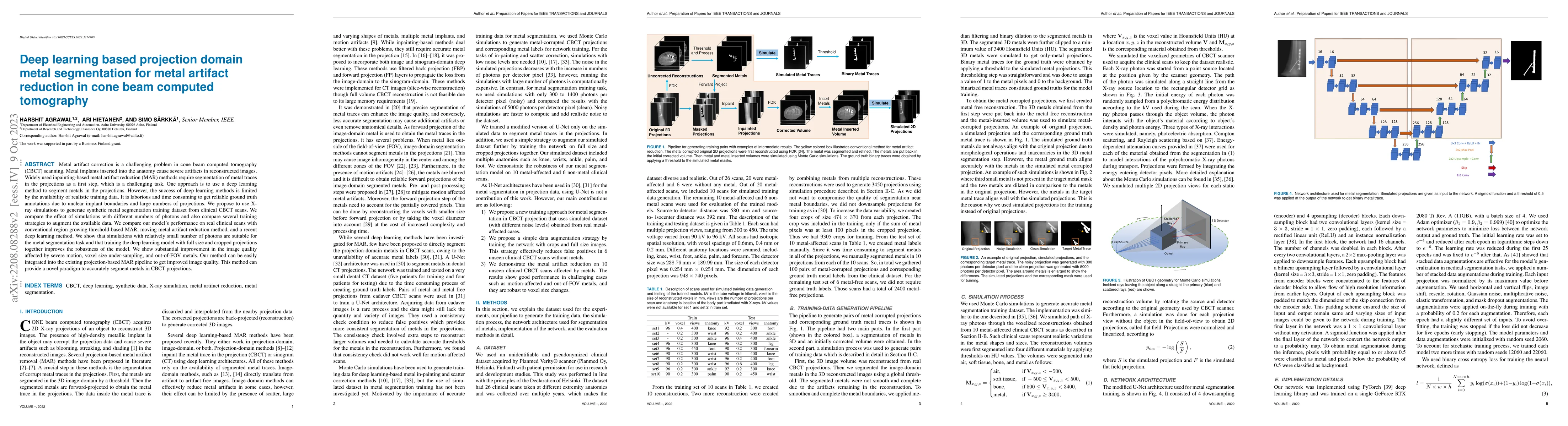
Metal artifact correction is a challenging problem in cone beam computed tomography (CBCT) scanning. Metal implants inserted into the anatomy cause severe artifacts in reconstructed images. Widely used inpainting-based metal artifact reduction (MAR) methods require segmentation of metal traces in the projections as a first step, which is a challenging task. One approach is to use a deep learning method to segment metals in the projections. However, the success of deep learning methods is limited by the availability of realistic training data. It is laborious and time consuming to get reliable ground truth annotations due to unclear implant boundaries and large numbers of projections. We propose to use X-ray simulations to generate synthetic metal segmentation training dataset from clinical CBCT scans. We compare the effect of simulations with different numbers of photons and also compare several training strategies to augment the available data. We compare our model's performance on real clinical scans with conventional region growing threshold-based MAR, moving metal artifact reduction method, and a recent deep learning method. We show that simulations with relatively small number of photons are suitable for the metal segmentation task and that training the deep learning model with full size and cropped projections together improves the robustness of the model. We show substantial improvement in the image quality affected by severe motion, voxel size under-sampling, and out-of-FOV metals. Our method can be easily integrated into the existing projection-based MAR pipeline to get improved image quality. This method can provide a novel paradigm to accurately segment metals in CBCT projections.
AI Key Findings
Get AI-generated insights about this paper's methodology, results, significance, and more — seven facets brought into focus.
Impact
Paper Details
Authors
PDF Preview
Key Terms
Citation Network
Current paper (gray), citations (green), references (blue)
Display is limited for performance on very large graphs.
Discussion 0