Publication
Metrics
AI Quick Summary
This paper proposes a convolutional neural network-based approach for deep learning-based super-resolution fluorescence microscopy that can effectively train on small datasets, overcoming the limitations of traditional methods that require large training sets. The new model successfully achieves super-resolution from a dataset of only 750 images, demonstrating its potential for use in other biomedical imaging modalities where large datasets are difficult to obtain.
Paper Preview
Abstract
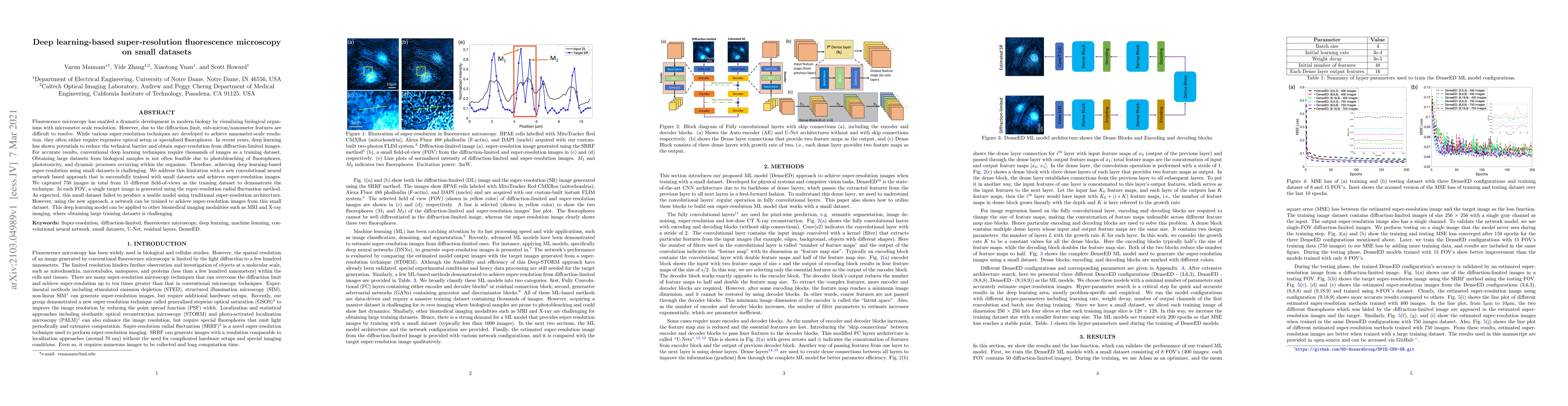
Fluorescence microscopy has enabled a dramatic development in modern biology by visualizing biological organisms with micrometer scale resolution. However, due to the diffraction limit, sub-micron/nanometer features are difficult to resolve. While various super-resolution techniques are developed to achieve nanometer-scale resolution, they often either require expensive optical setup or specialized fluorophores. In recent years, deep learning has shown the potentials to reduce the technical barrier and obtain super-resolution from diffraction-limited images. For accurate results, conventional deep learning techniques require thousands of images as a training dataset. Obtaining large datasets from biological samples is not often feasible due to the photobleaching of fluorophores, phototoxicity, and dynamic processes occurring within the organism. Therefore, achieving deep learning-based super-resolution using small datasets is challenging. We address this limitation with a new convolutional neural network-based approach that is successfully trained with small datasets and achieves super-resolution images. We captured 750 images in total from 15 different field-of-views as the training dataset to demonstrate the technique. In each FOV, a single target image is generated using the super-resolution radial fluctuation method. As expected, this small dataset failed to produce a usable model using traditional super-resolution architecture. However, using the new approach, a network can be trained to achieve super-resolution images from this small dataset. This deep learning model can be applied to other biomedical imaging modalities such as MRI and X-ray imaging, where obtaining large training datasets is challenging.
AI Key Findings
Get AI-generated insights about this paper's methodology, results, significance, and more — seven facets brought into focus.
Impact
Paper Details
Authors
PDF Preview
Key Terms
Citation Network
Current paper (gray), citations (green), references (blue)
Display is limited for performance on very large graphs.
Discussion 0