Deep learning enhanced mobile-phone microscopy
Publication
Metrics
AI Quick Summary
Researchers used deep learning to correct distortions in images taken with mobile phones for microscopy, producing high-resolution images comparable to laboratory-grade instruments. This method offers an affordable alternative for imaging applications and could enable standardization of optical images for clinical use.
Paper Preview
Abstract
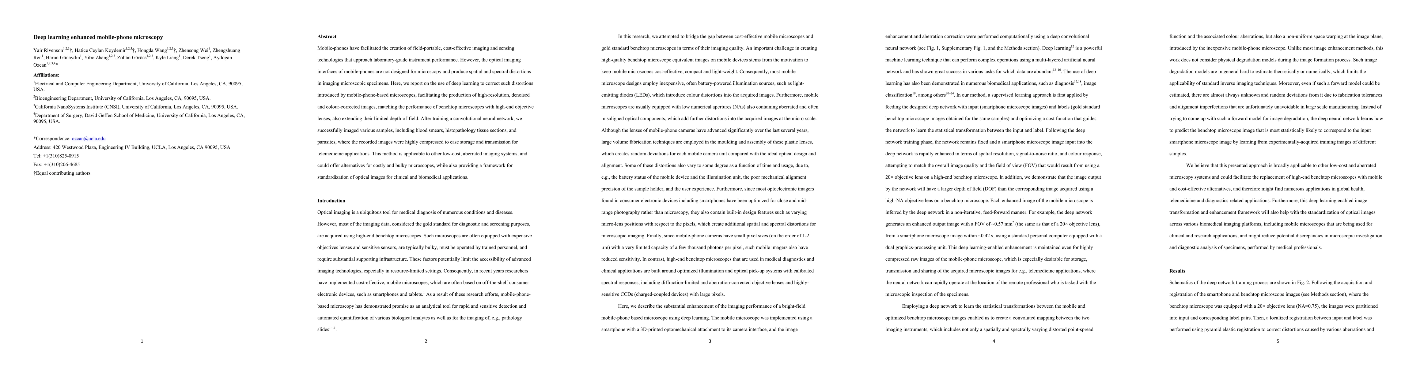
Mobile-phones have facilitated the creation of field-portable, cost-effective imaging and sensing technologies that approach laboratory-grade instrument performance. However, the optical imaging interfaces of mobile-phones are not designed for microscopy and produce spatial and spectral distortions in imaging microscopic specimens. Here, we report on the use of deep learning to correct such distortions introduced by mobile-phone-based microscopes, facilitating the production of high-resolution, denoised and colour-corrected images, matching the performance of benchtop microscopes with high-end objective lenses, also extending their limited depth-of-field. After training a convolutional neural network, we successfully imaged various samples, including blood smears, histopathology tissue sections, and parasites, where the recorded images were highly compressed to ease storage and transmission for telemedicine applications. This method is applicable to other low-cost, aberrated imaging systems, and could offer alternatives for costly and bulky microscopes, while also providing a framework for standardization of optical images for clinical and biomedical applications.
AI Key Findings
Get AI-generated insights about this paper's methodology, results, significance, and more — seven facets brought into focus.
Impact
Paper Details
PDF Preview
Key Terms
Citation Network
Current paper (gray), citations (green), references (blue)
Display is limited for performance on very large graphs.
Discussion 0