ArxivLens

Quick Summary:

This paper explores the use of deep learning for automatic spleen length measurement in sickle cell disease patients, aiming to reduce variability and improve accuracy compared to manual methods. The best model achieved a 7.42% error rate, nearing the inter-observer variability, marking the first fully automated approach for this task.

arXiv Id: 2009.02704
Comments: 9 pages, 2 figures
Date Published: 2020-09-08
Date Updated: 2020-09-08

Key Terms (TF-IDF):

ultrasound (0.333)
observer (0.247)
manual (0.243)
length (0.235)
observer (0.229)
length (0.225)
manual (0.224)
segmentation (0.215)
variability (0.209)
inter (0.204)
variability (0.197)
segmentation (0.195)
inter (0.190)
best (0.177)
estimation (0.173)
measurement (0.171)
images (0.165)
measurement (0.158)
estimation (0.158)
expertise (0.157)
License: http://arxiv.org/licenses/nonexclusive-distrib/1.0/

Summary:

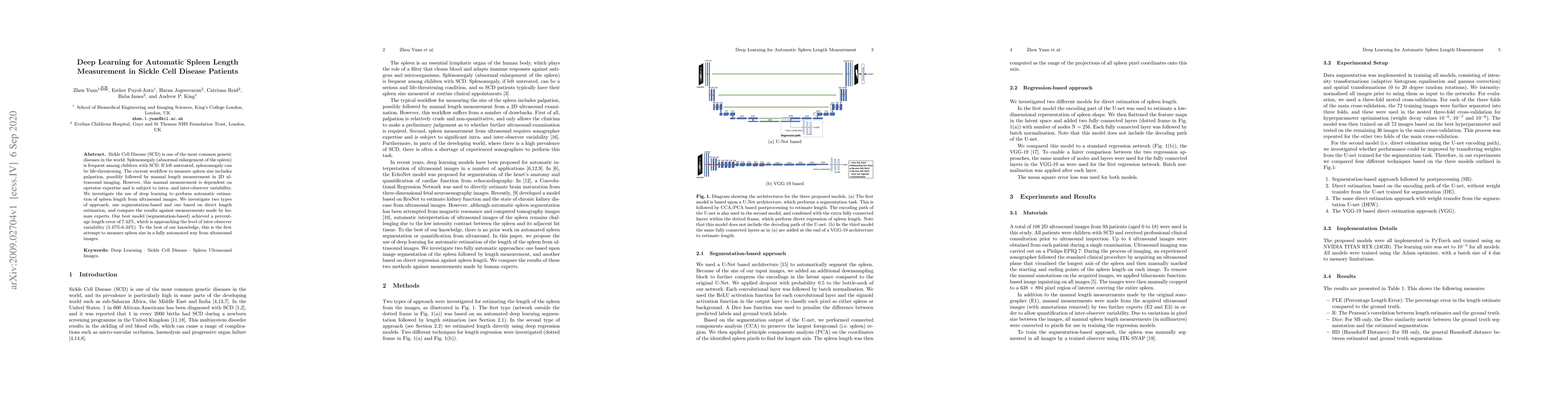
Sickle Cell Disease (SCD) is one of the most common genetic diseases in the world. Splenomegaly (abnormal enlargement of the spleen) is frequent among children with SCD. If left untreated, splenomegaly can be life-threatening. The current workflow to measure spleen size includes palpation, possibly followed by manual length measurement in 2D ultrasound imaging. However, this manual measurement is dependent on operator expertise and is subject to intra- and inter-observer variability. We investigate the use of deep learning to perform automatic estimation of spleen length from ultrasound images. We investigate two types of approach, one segmentation-based and one based on direct length estimation, and compare the results against measurements made by human experts. Our best model (segmentation-based) achieved a percentage length error of 7.42%, which is approaching the level of inter-observer variability (5.47%-6.34%). To the best of our knowledge, this is the first attempt to measure spleen size in a fully automated way from ultrasound images.

Citation & Reference Network Visualization

This graph shows how this paper is connected to other research. The current paper is shown in dark gray, papers that cite it are in green, and papers it references are in blue. You can:

Current Paper
Papers citing this work (Citations)
Papers cited by this work (References)

• Hover over any node to see the paper title

• Click a node to view that paper's details

• Drag nodes to rearrange the graph

• Use the zoom controls (bottom right) to zoom in/out

Citations and References

Title Authors Year Actions
DeepSPV: An Interpretable Deep Learning Pipeline for 3D Spleen Volume Estimation from 2D Ultrasound Images
Z. Yuan, D. Stojanovski, Lei Li, Alberto Gomez, Haran Jogeesvaran, Esther Puyol-Ant'on, B. Inusa, Andrew P. King
2024
View Paper
Application of Deep Learning for Real-Time Ablation Zone Measurement in Ultrasound Imaging
C. Zimmermann, Adrian Michelmann, Yannick Daniel, Markus D. Enderle, N. Salkić, W. Linzenbold
2024
View Paper
Deep Learning Framework for Spleen Volume Estimation from 2D Cross-sectional Views
Z. Yuan, E. Puyol-Antón, Haran Jogeesvaran, B. Inusa, A. King
2023
View Paper

Similar Papers

A Novel Deep Learning based Model for Erythrocytes Classification and Quantification in Sickle Cell Disease

Ajay Kumar, Prayag Tiwari, Pekka Marttinen, Vipin Kumar Rathi, Amit Kumar Jaiswal, Manish Bhatia, Balram Meena, Shagaf M Ansari

Published: 2023-05-04

DeepSPV: An Interpretable Deep Learning Pipeline for 3D Spleen Volume Estimation from 2D Ultrasound Images

Lei Li, Andrew P. King, Alberto Gomez, Zhen Yuan, Esther Puyol-Antón, David Stojanovski, Haran Jogeesvaran, Baba Inusa

Published: 2024-11-17

Automated Artifact Detection in Ultra-widefield Fundus Photography of Patients with Sickle Cell Disease

Mathias Unberath, Craig Jones, Anqi Feng, Dimitri Johnson, Grace R. Reilly, Loka Thangamathesvaran, Ann Nampomba, Adrienne W. Scott

Published: 2023-07-13

Comments (0)

This website uses cookies to ensure you get the best experience. Learn more