Authors
Summary
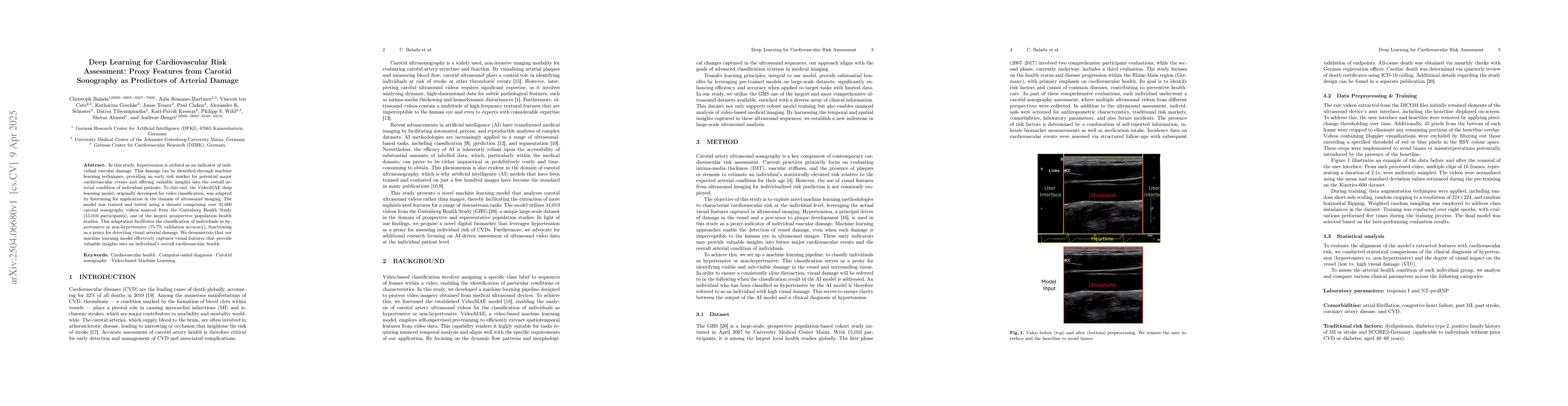
In this study, hypertension is utilized as an indicator of individual vascular damage. This damage can be identified through machine learning techniques, providing an early risk marker for potential major cardiovascular events and offering valuable insights into the overall arterial condition of individual patients. To this end, the VideoMAE deep learning model, originally developed for video classification, was adapted by finetuning for application in the domain of ultrasound imaging. The model was trained and tested using a dataset comprising over 31,000 carotid sonography videos sourced from the Gutenberg Health Study (15,010 participants), one of the largest prospective population health studies. This adaptation facilitates the classification of individuals as hypertensive or non-hypertensive (75.7% validation accuracy), functioning as a proxy for detecting visual arterial damage. We demonstrate that our machine learning model effectively captures visual features that provide valuable insights into an individual's overall cardiovascular health.
AI Key Findings
Generated Jun 09, 2025
Methodology
This study adapted the VideoMAE deep learning model, originally for video classification, for analyzing carotid sonography videos to classify individuals as hypertensive or non-hypertensive, serving as a proxy for detecting visual arterial damage. The model was trained and validated using over 31,000 videos from the Gutenberg Health Study (GHS) dataset, comprising 15,010 participants.
Key Results
- The adapted VideoMAE model achieved a validation accuracy of 75.7% in classifying individuals as hypertensive or non-hypertensive.
- Individuals classified as having high visual damage, regardless of hypertension diagnosis, showed significantly worse cardiovascular health conditions compared to those with low visual damage.
- Non-hypertensive individuals with high visual damage exhibited a 1.9-fold higher likelihood of dyslipidemia and a 4.9-fold higher likelihood of type 2 diabetes mellitus compared to the baseline of non-hypertensive individuals with low visual damage.
Significance
This research demonstrates the potential of deep learning in identifying cardiovascular risk using carotid sonography, offering valuable insights into an individual's overall arterial condition and serving as an early risk marker for major cardiovascular events.
Technical Contribution
The study introduced the use of VideoMAE, a deep learning model originally for video classification, adapted for carotid sonography analysis, demonstrating its effectiveness in identifying cardiovascular risk markers.
Novelty
This work is novel in its application of deep learning techniques to carotid sonography for cardiovascular risk assessment, providing a non-invasive, cost-effective method for evaluating arterial damage as a proxy for hypertension and subsequent cardiovascular events.
Limitations
- The study relied on proxy features for hypertension classification due to the absence of direct blood pressure measurement via ultrasound.
- Performance dip observed in individuals aged 52 to 62 warrants further investigation into the underlying reasons.
Future Work
- Conduct a more detailed analysis of various clinical variables to evaluate their utility as potential risk prediction proxies.
- Develop an innovative, patient-centered approach for risk assessment by integrating multiple proxy classifiers.
Paper Details
PDF Preview
Citation Network
Current paper (gray), citations (green), references (blue)
Display is limited for performance on very large graphs.
Similar Papers
Found 4 papersCarotid and femoral bifurcation plaques detected by ultrasound as predictors of cardiovascular events.
Heiss, Christian, Rammos, Christos, Paraskevas, Kosmas I et al.
Deep Learning-based Prediction of Stress and Strain Maps in Arterial Walls for Improved Cardiovascular Risk Assessment
Xianqi Li, Linxia Gu, Yasin Shokrollahi1 et al.
No citations found for this paper.
Comments (0)