Deep Learning Models Delineates Multiple Nuclear Phenotypes in H&E Stained Histology Sections
Publication
Metrics
AI Quick Summary
Deep learning models, specifically fused very deep convolutional networks, successfully segment complex nuclear phenotypes in histology sections, overcoming challenges like nuclear geometry variations and overlapping nuclei. The method was validated on diverse breast and brain histology samples.
Paper Preview
Abstract
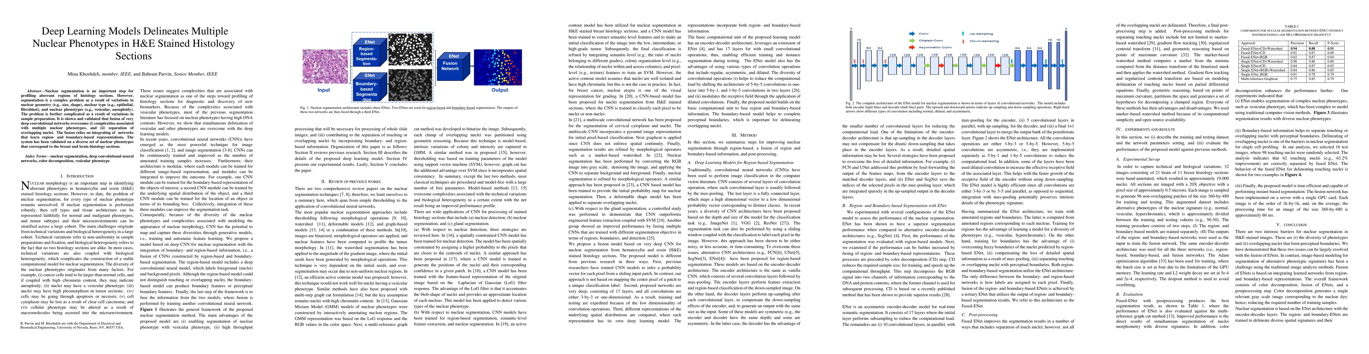
Nuclear segmentation is an important step for profiling aberrant regions of histology sections. However, segmentation is a complex problem as a result of variations in nuclear geometry (e.g., size, shape), nuclear type (e.g., epithelial, fibroblast), and nuclear phenotypes (e.g., vesicular, aneuploidy). The problem is further complicated as a result of variations in sample preparation. It is shown and validated that fusion of very deep convolutional networks overcomes (i) complexities associated with multiple nuclear phenotypes, and (ii) separation of overlapping nuclei. The fusion relies on integrating of networks that learn region- and boundary-based representations. The system has been validated on a diverse set of nuclear phenotypes that correspond to the breast and brain histology sections.
AI Key Findings
Get AI-generated insights about this paper's methodology, results, significance, and more — seven facets brought into focus.
Impact
Paper Details
PDF Preview
Key Terms
Citation Network
Current paper (gray), citations (green), references (blue)
Display is limited for performance on very large graphs.
Discussion 0