Publication
Metrics
AI Quick Summary
This paper proposes a deep learning model for brain tumor segmentation using both fully-annotated and weakly-annotated images, combining segmentation and image-level classification to improve performance. The method shows significant improvement over standard supervised learning, especially when more weakly-annotated data is available.
Paper Preview
Abstract
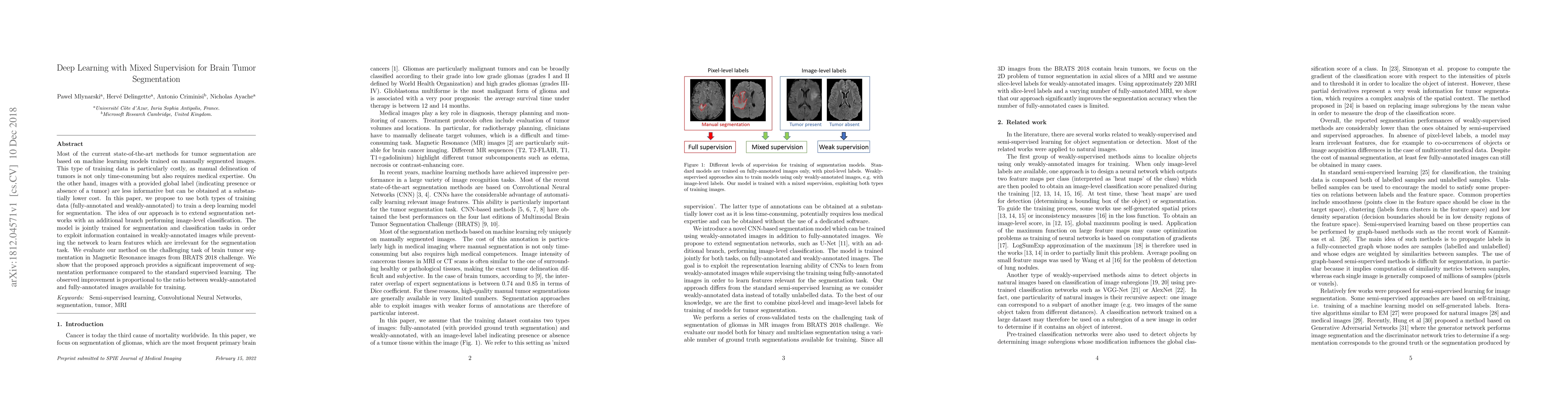
Most of the current state-of-the-art methods for tumor segmentation are based on machine learning models trained on manually segmented images. This type of training data is particularly costly, as manual delineation of tumors is not only time-consuming but also requires medical expertise. On the other hand, images with a provided global label (indicating presence or absence of a tumor) are less informative but can be obtained at a substantially lower cost. In this paper, we propose to use both types of training data (fully-annotated and weakly-annotated) to train a deep learning model for segmentation. The idea of our approach is to extend segmentation networks with an additional branch performing image-level classification. The model is jointly trained for segmentation and classification tasks in order to exploit information contained in weakly-annotated images while preventing the network to learn features which are irrelevant for the segmentation task. We evaluate our method on the challenging task of brain tumor segmentation in Magnetic Resonance images from BRATS 2018 challenge. We show that the proposed approach provides a significant improvement of segmentation performance compared to the standard supervised learning. The observed improvement is proportional to the ratio between weakly-annotated and fully-annotated images available for training.
AI Key Findings
Get AI-generated insights about this paper's methodology, results, significance, and more — seven facets brought into focus.
Impact
Paper Details
Authors
PDF Preview
Key Terms
Citation Network
Current paper (gray), citations (green), references (blue)
Display is limited for performance on very large graphs.
Discussion 0