Deep Morphing: Detecting bone structures in fluoroscopic X-ray images with prior knowledge
Publication
Metrics
AI Quick Summary
This paper proposes a deep learning approach called 'Deep Morphing' to detect bone structures in low-quality fluoroscopic X-ray images with limited training data, using simple geometrical models or statistical shape representations.
Paper Preview
Abstract
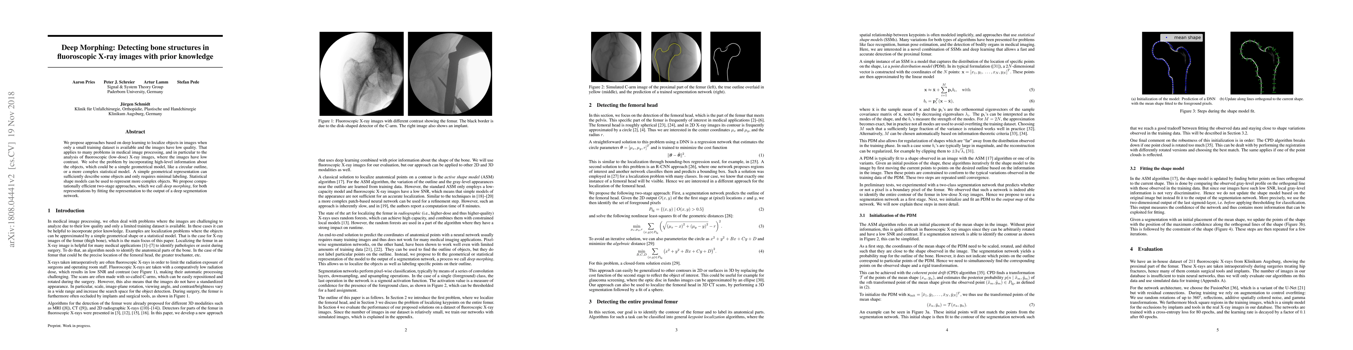
We propose approaches based on deep learning to localize objects in images when only a small training dataset is available and the images have low quality. That applies to many problems in medical image processing, and in particular to the analysis of fluoroscopic (low-dose) X-ray images, where the images have low contrast. We solve the problem by incorporating high-level information about the objects, which could be a simple geometrical model, like a circular outline, or a more complex statistical model. A simple geometrical representation can sufficiently describe some objects and only requires minimal labeling. Statistical shape models can be used to represent more complex objects. We propose computationally efficient two-stage approaches, which we call deep morphing, for both representations by fitting the representation to the output of a deep segmentation network.
AI Key Findings
Get AI-generated insights about this paper's methodology, results, significance, and more — seven facets brought into focus.
Impact
Paper Details
PDF Preview
Key Terms
Citation Network
Current paper (gray), citations (green), references (blue)
Display is limited for performance on very large graphs.
Discussion 0